
Search Count: 82
 |
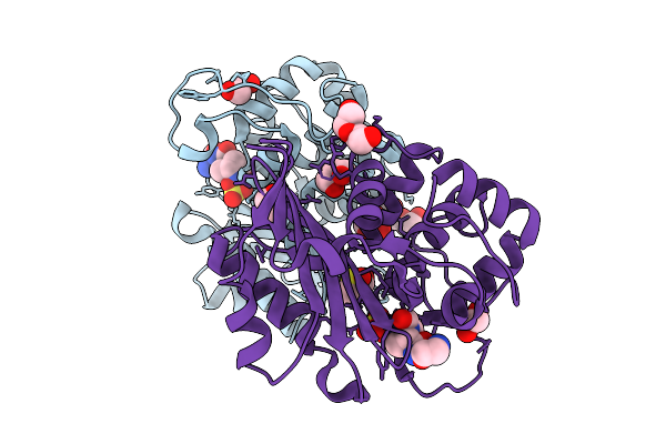
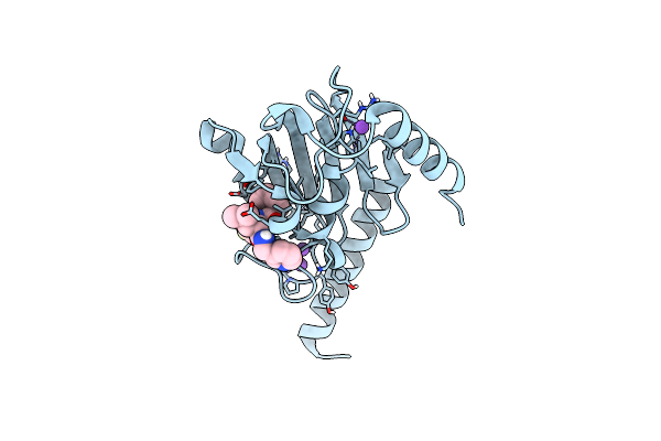
Organism: Klebsiella pneumoniae
Method: X-RAY DIFFRACTION Release Date: 2025-09-03 Classification: ANTIMICROBIAL PROTEIN Ligands: CL, GOL |
 |
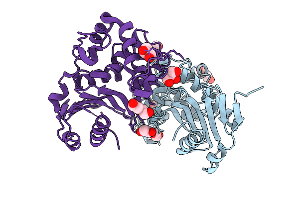
Organism: Klebsiella pneumoniae
Method: X-RAY DIFFRACTION Release Date: 2025-09-03 Classification: ANTIMICROBIAL PROTEIN Ligands: CL, PGE, PEG, EDO, OP0, DMS, GOL, PG4 |
 |
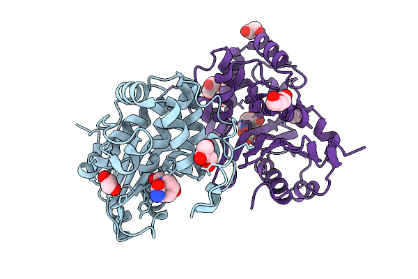
Organism: Enterobacter cloacae
Method: X-RAY DIFFRACTION Release Date: 2025-09-03 Classification: ANTIMICROBIAL PROTEIN Ligands: CL, GOL, EDO, PEG |
 |
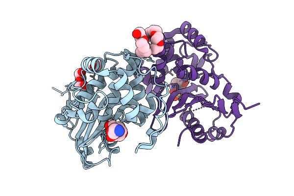
Organism: Enterobacter cloacae
Method: X-RAY DIFFRACTION Release Date: 2025-09-03 Classification: ANTIMICROBIAL PROTEIN Ligands: NXL, A1IY4, CL, EDO, 2PE |
 |
Organism: Enterobacter cloacae
Method: X-RAY DIFFRACTION Release Date: 2025-09-03 Classification: ANTIMICROBIAL PROTEIN Ligands: OP0, EDO, GOL, PEG, CL, DMS |
 |
Organism: Serratia marcescens
Method: X-RAY DIFFRACTION Release Date: 2025-09-03 Classification: ANTIMICROBIAL PROTEIN Ligands: PEG, CL, GOL, PG4, EDO |
 |
Organism: Serratia marcescens
Method: X-RAY DIFFRACTION Release Date: 2025-09-03 Classification: ANTIMICROBIAL PROTEIN Ligands: GOL, EDO, NXL, A1IY4, CL, PGE, PEG |
 |
Organism: Serratia marcescens
Method: X-RAY DIFFRACTION Release Date: 2025-09-03 Classification: ANTIMICROBIAL PROTEIN Ligands: OP0, PEG, EDO, A1IYS, CL, 2PE |
 |
Organism: Enterobacter cloacae
Method: X-RAY DIFFRACTION Release Date: 2025-09-03 Classification: ANTIMICROBIAL PROTEIN Ligands: OP0, EDO, GOL, A1IYS, CL, 2PE |
 |
Organism: Coxiella burnetii
Method: X-RAY DIFFRACTION Release Date: 2025-08-13 Classification: CYTOSOLIC PROTEIN Ligands: EPE, EDO, PEG, CL |
 |
Crystal Structure Of The Coxiella Burnetii E110Q Mutant 2-Methylisocitrate Lyase
Organism: Coxiella burnetii
Method: X-RAY DIFFRACTION Release Date: 2025-08-13 Classification: CYTOSOLIC PROTEIN Ligands: EDO, CL |
 |
Crystal Structure Of The Coxiella Burnetii 2-Methylisocitrate Lyase Bound To Substrate 2-Mic
Organism: Coxiella burnetii
Method: X-RAY DIFFRACTION Release Date: 2025-08-13 Classification: CYTOSOLIC PROTEIN Ligands: MIC, EOH, EDO, MG, CL |
 |
Crystal Structure Of The Coxiella Burnetii R152Q Mutant 2-Methylisocitrate Lyase
Organism: Coxiella burnetii
Method: X-RAY DIFFRACTION Release Date: 2025-08-13 Classification: CYTOSOLIC PROTEIN Ligands: CIT, EDO, CL |
 |
Crystal Structure Of The Coxiella Burnetii 2-Methylisocitrate Lyase Bound To Inhibitor Isocitric Acid
Organism: Coxiella burnetii
Method: X-RAY DIFFRACTION Release Date: 2025-08-13 Classification: CYTOSOLIC PROTEIN Ligands: ICT, EDO, MG, CL, PEG, PGE |
 |
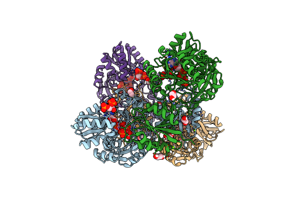
Crystal Structure Of The Coxiella Burnetii 2-Methylisocitrate Lyase Bound To Products Succinic And Pyruvic Acid
Organism: Coxiella burnetii
Method: X-RAY DIFFRACTION Release Date: 2025-08-13 Classification: CYTOSOLIC PROTEIN Ligands: SIN, PYR, EDO, MG, CL, DMS, PGE, PEG |
 |
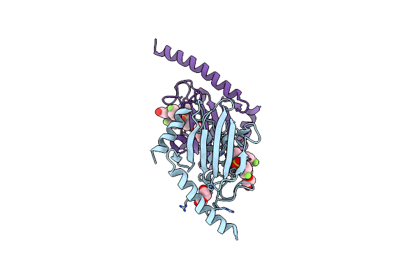
Organism: Burkholderia pseudomallei
Method: X-RAY DIFFRACTION Resolution:2.02 Å Release Date: 2024-06-12 Classification: STRUCTURAL PROTEIN Ligands: WRX, GOL, PEG |
 |
Organism: Trypanosoma cruzi
Method: X-RAY DIFFRACTION Resolution:1.71 Å Release Date: 2024-06-12 Classification: STRUCTURAL PROTEIN Ligands: WS5, NA |
 |
Organism: Trypanosoma cruzi
Method: X-RAY DIFFRACTION Resolution:2.64 Å Release Date: 2024-06-12 Classification: STRUCTURAL PROTEIN Ligands: WRX, PEG |
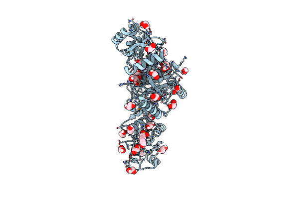 |
Cyclic 2,3-Diphosphoglycerate Synthetase From The Hyperthermophilic Archaeon Methanothermus Fervidus
Organism: Methanothermus fervidus dsm 2088
Method: X-RAY DIFFRACTION Resolution:1.64 Å Release Date: 2023-12-06 Classification: LIGASE Ligands: MPO, EDO, PEG, NA, CL |
 |
Cyclic 2,3-Diphosphoglycerate Synthetase From The Hyperthermophilic Archaeon Methanothermus Fervidus Bound To 2,3-Diphosphoglycerate And Adp.
Organism: Methanothermus fervidus dsm 2088
Method: X-RAY DIFFRACTION Resolution:2.23 Å Release Date: 2023-12-06 Classification: LIGASE Ligands: ADP, EDO, DG2, MG, PO4 |