
Search Count: 15
All
Selected
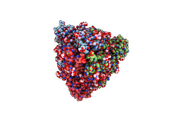 |
Organism: Severe acute respiratory syndrome coronavirus 2, Bat coronavirus rp3/2004, Homo sapiens
Method: ELECTRON MICROSCOPY Release Date: 2022-02-23 Classification: VIRAL PROTEIN Ligands: NAG |
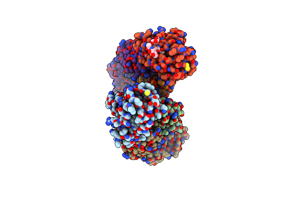 |
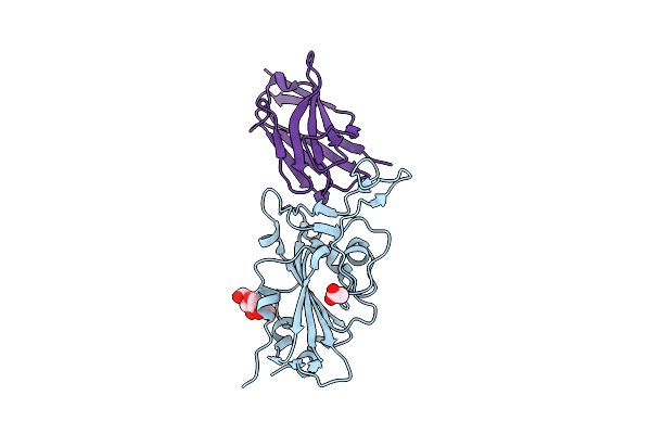
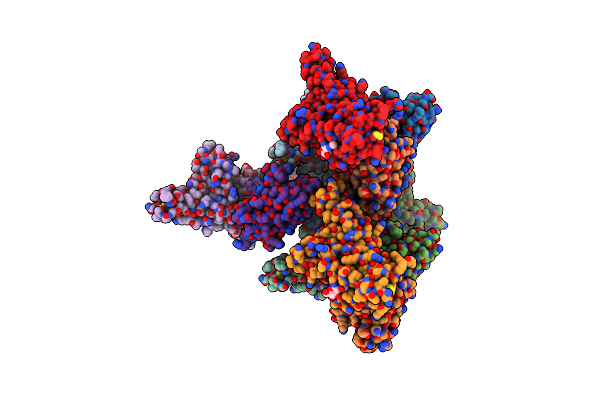
Crystal Structure Of The Receptor Binding Domain Of Sars-Cov-2 Spike Glycoprotein In Complex With Fi-3A Fab
Organism: Severe acute respiratory syndrome coronavirus 2, Homo sapiens
Method: X-RAY DIFFRACTION Resolution:3.00 Å Release Date: 2022-02-02 Classification: VIRAL PROTEIN |
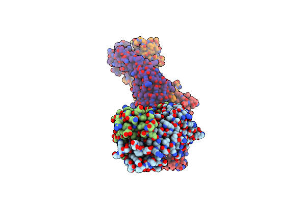 |
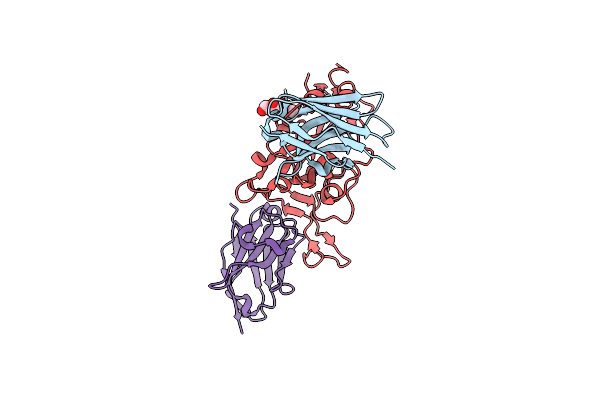
Crystal Structure Of The Receptor Binding Domain Of Sars-Cov-2 Spike Glycoprotein In Complex With Fi-3A And Fd-11A Fabs
Organism: Severe acute respiratory syndrome coronavirus 2, Homo sapiens
Method: X-RAY DIFFRACTION Resolution:3.20 Å Release Date: 2022-02-02 Classification: VIRAL PROTEIN Ligands: PO4 |
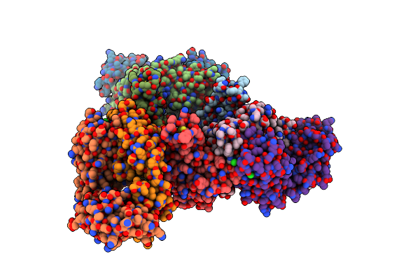 |
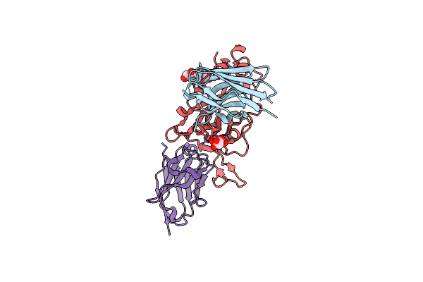
Crystal Structure Of The Receptor Binding Domain Of Sars-Cov-2 Spike Glycoprotein In Complex With Fd-5D Fab
Organism: Severe acute respiratory syndrome coronavirus 2, Homo sapiens
Method: X-RAY DIFFRACTION Resolution:2.92 Å Release Date: 2022-02-02 Classification: VIRAL PROTEIN Ligands: NAG, SO4, NO3, CL, GOL |
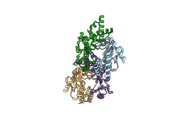 |
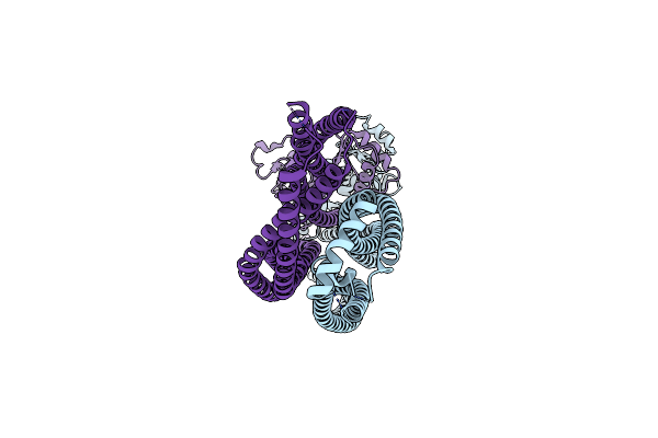
Crystal Structure Of Mouse Smc1-Smc3 Hinge Domain Containing A D574Y Mutation
Organism: Mus musculus
Method: X-RAY DIFFRACTION Resolution:2.00 Å Release Date: 2021-11-17 Classification: DNA BINDING PROTEIN |
 |
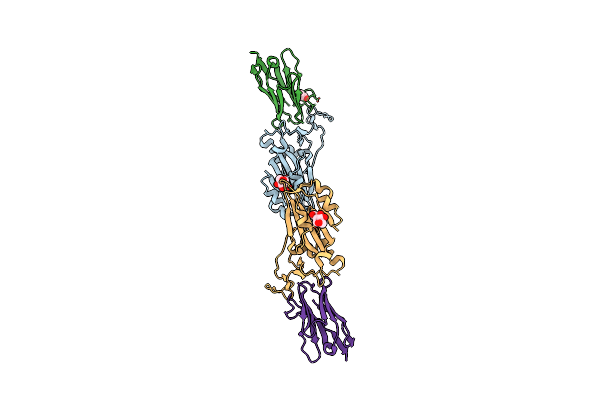
Crystal Structure Of Sars-Cov-2 Spike Protein Receptor-Binding Domain Complexed With A Pre-Pandemic Antibody S-B8 Fab
Organism: Severe acute respiratory syndrome coronavirus 2, Homo sapiens
Method: X-RAY DIFFRACTION Resolution:2.25 Å Release Date: 2021-09-22 Classification: VIRAL PROTEIN/IMMUNE SYSTEM Ligands: NAG, EDO, SO4, PGE |
 |
Crystal Structure Of Sars-Cov-2 Spike Protein Receptor-Binding Domain Complexed With A Pre-Pandemic Antibody S-E6 Fab
Organism: Severe acute respiratory syndrome coronavirus 2, Homo sapiens
Method: X-RAY DIFFRACTION Resolution:2.70 Å Release Date: 2021-09-22 Classification: VIRAL PROTEIN/IMMUNE SYSTEM Ligands: NAG |
 |
Organism: Severe acute respiratory syndrome coronavirus 2, Vicugna pacos
Method: ELECTRON MICROSCOPY Resolution:3.00 Å Release Date: 2021-08-11 Classification: ANTIVIRAL PROTEIN Ligands: NAG |
 |
Organism: Severe acute respiratory syndrome coronavirus 2, Lama glama
Method: X-RAY DIFFRACTION Resolution:1.50 Å Release Date: 2021-08-11 Classification: ANTIVIRAL PROTEIN Ligands: NAG, ACT |
 |
Organism: Lama glama, Severe acute respiratory syndrome coronavirus 2
Method: X-RAY DIFFRACTION Resolution:1.90 Å Release Date: 2021-08-11 Classification: ANTIVIRAL PROTEIN Ligands: NAG, CIT, CL |
 |
Organism: Lama glama, Severe acute respiratory syndrome coronavirus 2
Method: X-RAY DIFFRACTION Resolution:1.55 Å Release Date: 2021-08-11 Classification: ANTIVIRAL PROTEIN Ligands: NAG, CIT, CL |
 |
Organism: Severe acute respiratory syndrome coronavirus 2, Lama glama
Method: X-RAY DIFFRACTION Resolution:1.65 Å Release Date: 2021-08-11 Classification: ANTIVIRAL PROTEIN Ligands: NAG, GOL |
 |
Organism: Severe acute respiratory syndrome coronavirus 2, Lama glama
Method: X-RAY DIFFRACTION Resolution:2.34 Å Release Date: 2021-08-11 Classification: ANTIVIRAL PROTEIN Ligands: NAG |
 |
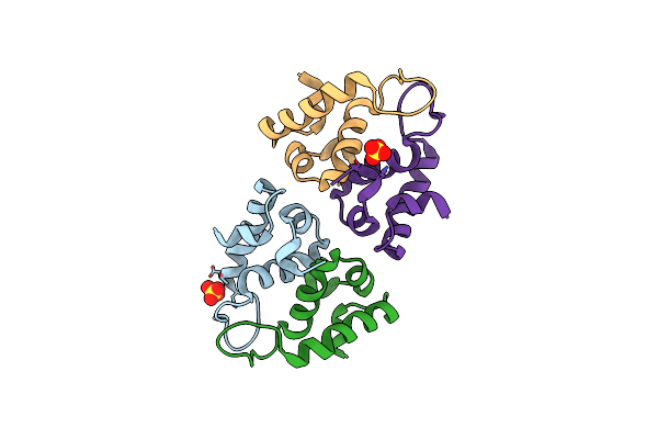
Organism: Saccharomyces cerevisiae (strain atcc 204508 / s288c)
Method: ELECTRON MICROSCOPY Release Date: 2021-07-28 Classification: CELL CYCLE |
 |
Organism: Escherichia coli
Method: X-RAY DIFFRACTION Resolution:1.50 Å Release Date: 2011-08-24 Classification: UNKNOWN FUNCTION Ligands: SO4 |