
Search Count: 13
 |
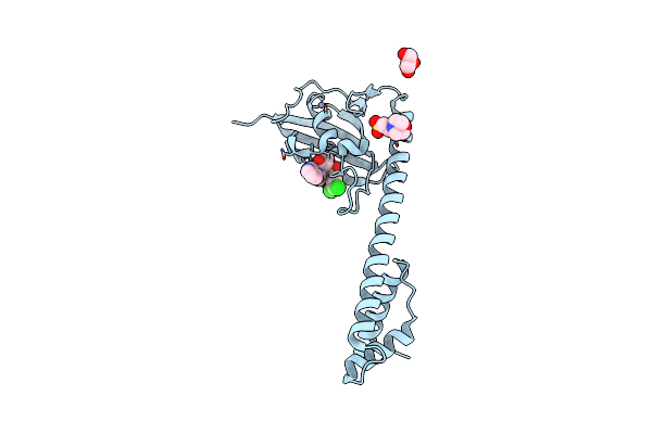
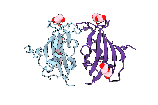
Organism: Burkholderia pseudomallei
Method: X-RAY DIFFRACTION Resolution:2.02 Å Release Date: 2024-06-12 Classification: STRUCTURAL PROTEIN Ligands: WRX, GOL, PEG |
 |
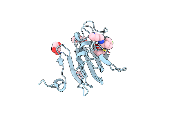
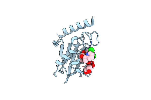
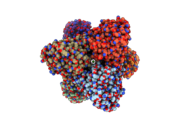
Organism: Trypanosoma cruzi
Method: X-RAY DIFFRACTION Resolution:1.71 Å Release Date: 2024-06-12 Classification: STRUCTURAL PROTEIN Ligands: WS5, NA |
 |
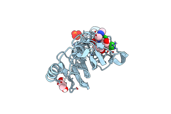
Organism: Trypanosoma cruzi
Method: X-RAY DIFFRACTION Resolution:2.64 Å Release Date: 2024-06-12 Classification: STRUCTURAL PROTEIN Ligands: WRX, PEG |
 |
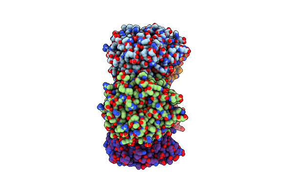
Organism: Legionella pneumophila
Method: X-RAY DIFFRACTION Resolution:1.71 Å Release Date: 2023-09-06 Classification: STRUCTURAL PROTEIN Ligands: GOL, EPE, MES, ZN |
 |
Organism: Legionella pneumophila
Method: X-RAY DIFFRACTION Resolution:2.40 Å Release Date: 2023-09-06 Classification: STRUCTURAL PROTEIN Ligands: 9QN, MES, ZN, GOL |
 |
Organism: Legionella pneumophila
Method: X-RAY DIFFRACTION Resolution:1.49 Å Release Date: 2023-09-06 Classification: STRUCTURAL PROTEIN Ligands: GOL, WRL |
 |
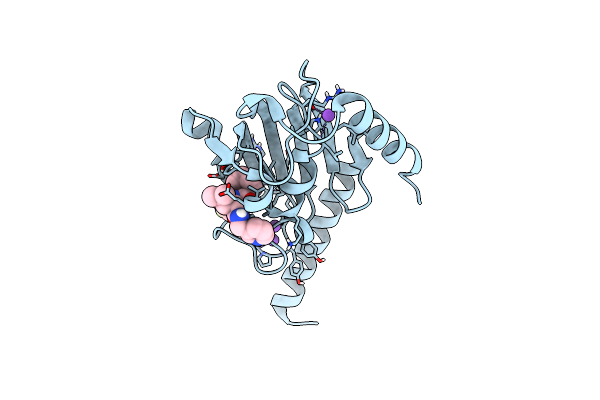
Organism: Trypanosoma cruzi
Method: X-RAY DIFFRACTION Resolution:1.34 Å Release Date: 2023-09-06 Classification: STRUCTURAL PROTEIN Ligands: GOL, WRL, PO4 |
 |
Organism: Legionella pneumophila
Method: X-RAY DIFFRACTION Resolution:1.44 Å Release Date: 2023-09-06 Classification: STRUCTURAL PROTEIN Ligands: 9QN, NA |
 |
Organism: Legionella pneumophila
Method: X-RAY DIFFRACTION Resolution:2.26 Å Release Date: 2023-09-06 Classification: STRUCTURAL PROTEIN Ligands: PEG, MES |
 |
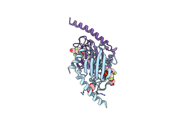
Organism: Formosa sp. hel1_33_131
Method: X-RAY DIFFRACTION Resolution:2.60 Å Release Date: 2018-12-26 Classification: HYDROLASE Ligands: CA |
 |
Organism: Castellaniella defragrans
Method: X-RAY DIFFRACTION Resolution:1.91 Å Release Date: 2016-04-27 Classification: ISOMERASE Ligands: PG0 |
 |
Organism: Castellaniella defragrans
Method: X-RAY DIFFRACTION Resolution:2.50 Å Release Date: 2016-04-27 Classification: LYASE Ligands: PG0, 64Z, 650 |
 |
The Crystal Structure Of Fad And Thdp Dependent Cyclohexane-1,2-Dione Hydrolase (Cdh) From Azoarcus Sp. Strain 22Lin
Organism: Azoarcus sp.
Method: X-RAY DIFFRACTION Resolution:1.26 Å Release Date: 2008-04-22 Classification: HYDROLASE Ligands: MG, CL, FAD, TPP, MPD, PO4 |