
Search Count: 7
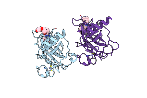 |
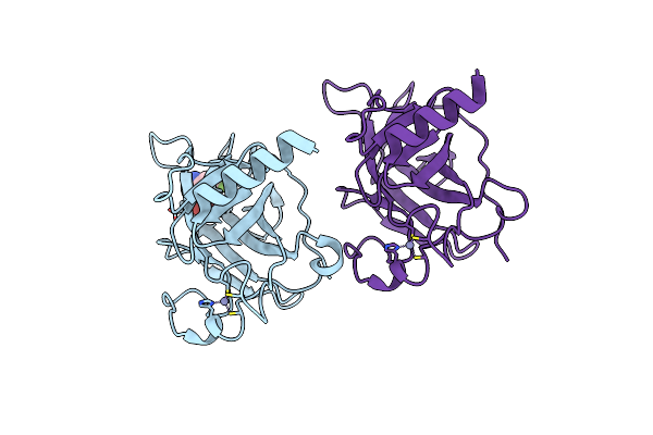
Structure Of The P53 Cancer Mutant Y220C With Bound Small Molecule 7- Ethyl-3-(Piperidin-4-Yl)-1H-Indole
Organism: Homo sapiens
Method: X-RAY DIFFRACTION Resolution:1.36 Å Release Date: 2015-12-16 Classification: TRANSCRIPTION Ligands: ZN, PEG, 92O |
 |
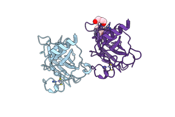
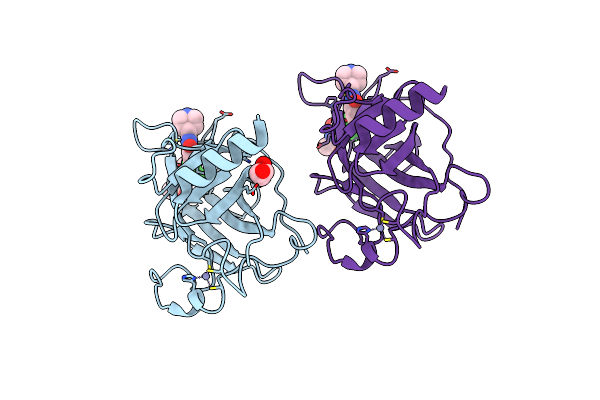
Structure Of The P53 Cancer Mutant Y220C With Bound Small-Molecule Stabilizer Phikan5149
Organism: Homo sapiens
Method: X-RAY DIFFRACTION Resolution:1.62 Å Release Date: 2015-12-16 Classification: TRANSCRIPTION Ligands: ZN, UL7 |
 |
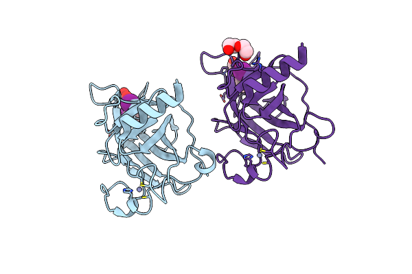
Structure Of The P53 Cancer Mutant Y220C In Complex With An Indole- Based Small Molecule
Organism: Homo sapiens
Method: X-RAY DIFFRACTION Resolution:1.78 Å Release Date: 2015-12-16 Classification: SIGNALING PROTEIN Ligands: ZN, PEG, RZH |
 |
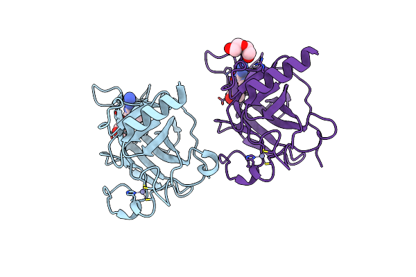
Structure Of The P53 Cancer Mutant Y220C In Complex With 2-Hydroxy-3, 5-Diiodo-4-(1H-Pyrrol-1-Yl)Benzoic Acid
Organism: Homo sapiens
Method: X-RAY DIFFRACTION Resolution:1.47 Å Release Date: 2015-12-16 Classification: CELL CYCLE Ligands: Y0V, ZN, PEG |
 |
Structure Of The P53 Cancer Mutant Y220C With Bound Small Molecule Phikan7099
Organism: Homo sapiens
Method: X-RAY DIFFRACTION Resolution:1.35 Å Release Date: 2015-12-16 Classification: CELL CYCLE Ligands: ZN, GOL, GOH, PEG |
 |
Structure Of The P53 Cancer Mutant Y220C With Bound 3-Bromo-5-(Trifluoromethyl)Benzene-1,2-Diamine
Organism: Homo sapiens
Method: X-RAY DIFFRACTION Resolution:1.50 Å Release Date: 2015-12-16 Classification: CELL CYCLE Ligands: UFV, ZN |
 |
Structure Of The P53 Cancer Mutant Y220C With Bound Small Molecule Phikan883
Organism: Homo sapiens
Method: X-RAY DIFFRACTION Resolution:1.74 Å Release Date: 2015-12-16 Classification: CELL CYCLE Ligands: FY8, GOL, ZN |