
Search Count: 2,574
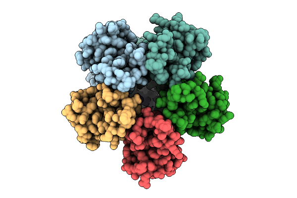 |
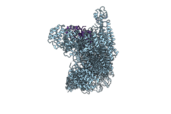
Organism: Escherichia coli
Method: ELECTRON MICROSCOPY Release Date: 2025-12-17 Classification: MEMBRANE PROTEIN |
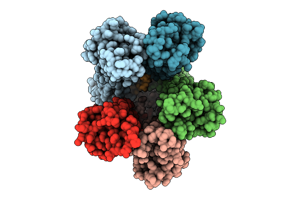 |
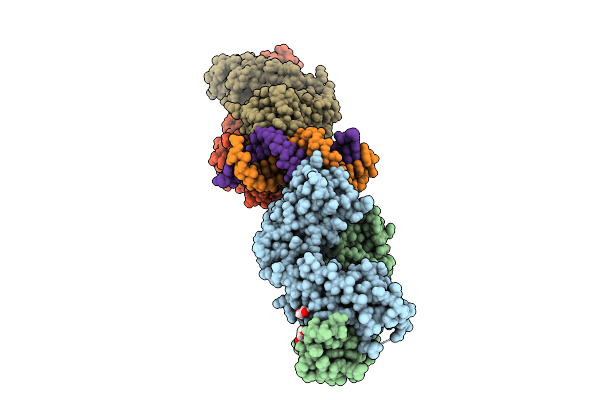
Organism: Escherichia coli str. k-12 substr. mg1655
Method: ELECTRON MICROSCOPY Release Date: 2025-12-17 Classification: MEMBRANE PROTEIN |
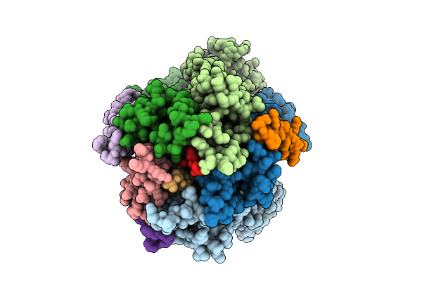 |
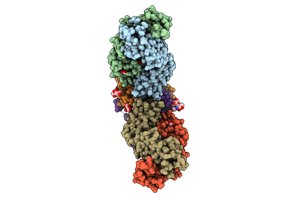
Organism: Escherichia coli str. k-12 substr. mg1655
Method: ELECTRON MICROSCOPY Release Date: 2025-12-17 Classification: MEMBRANE PROTEIN |
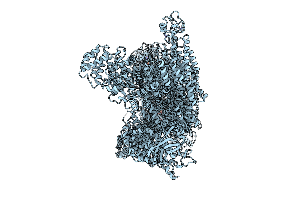 |
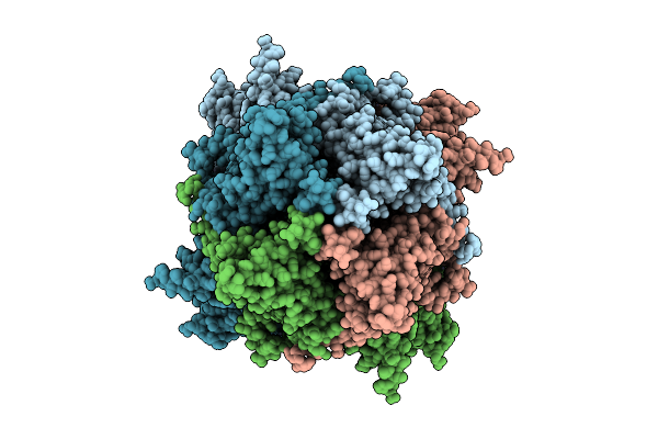
Organism: Mus musculus
Method: ELECTRON MICROSCOPY Release Date: 2025-12-10 Classification: LIGASE Ligands: ATP, MG, ZN |
 |
Cryo-Em Structure Of Mouse Rnf213:Ube2L3 Transthiolation Intermediate, Chemically Stabilized, And Atpgs
Organism: Mus musculus, Homo sapiens
Method: ELECTRON MICROSCOPY Release Date: 2025-12-10 Classification: LIGASE Ligands: ATP, MG, ZN, ADP |
 |
Organism: Mus musculus, Synthetic construct
Method: X-RAY DIFFRACTION Release Date: 2025-12-10 Classification: RNA BINDING PROTEIN/RNA Ligands: PEG |
 |
Organism: Homo sapiens
Method: X-RAY DIFFRACTION Release Date: 2025-12-10 Classification: SIGNALING PROTEIN Ligands: EDO, MAN, NAG, CIT |
 |
Structure-Guided Design Of Picomolar-Level Macrocyclic Trpc5 Channel Inhibitors With Antidepressant Activity
Organism: Mus musculus
Method: ELECTRON MICROSCOPY Release Date: 2025-12-03 Classification: MEMBRANE PROTEIN Ligands: ZN, CA, A1EH7 |
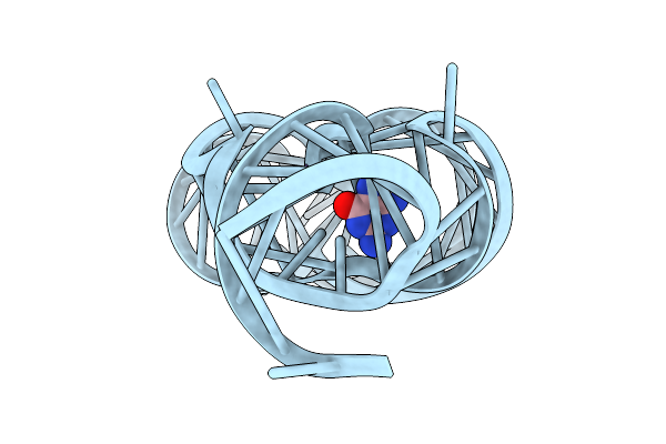 |
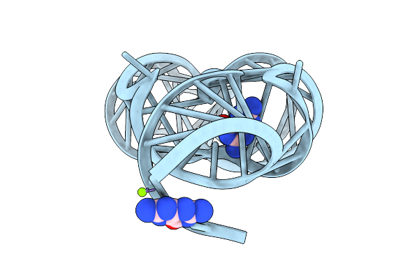
Organism: Paenibacillus sp. fsl e2-0178
Method: X-RAY DIFFRACTION Release Date: 2025-11-26 Classification: RNA Ligands: GUN, MG |
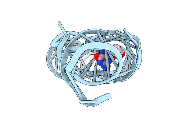 |
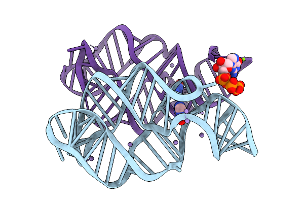
Crystal Structure Of Guanine-Ii Riboswitch In Complex With 2'-Deoxyguanosine
Organism: Paenibacillus sp. fsl e2-0178
Method: X-RAY DIFFRACTION Release Date: 2025-11-26 Classification: RNA Ligands: MG, GNG |
 |
Organism: Paenibacillus sp. fsl e2-0178
Method: X-RAY DIFFRACTION Release Date: 2025-11-26 Classification: RNA Ligands: HPA, GTP, MG |
 |
Organism: Paenibacillus sp. fsl e2-0178
Method: X-RAY DIFFRACTION Release Date: 2025-11-26 Classification: RNA Ligands: GMP, MG |
 |
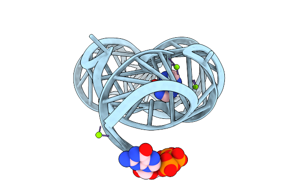
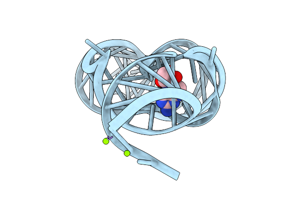
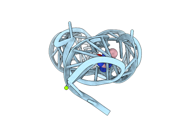
Crystal Structure Of Guanine-Ii Riboswitch In Complex With Guanine Soaked With Mn2+
Organism: Paenibacillus sp. fsl e2-0178
Method: X-RAY DIFFRACTION Release Date: 2025-11-26 Classification: RNA Ligands: GUN, MN, GTP, MG |
 |
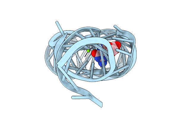
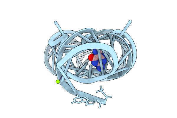
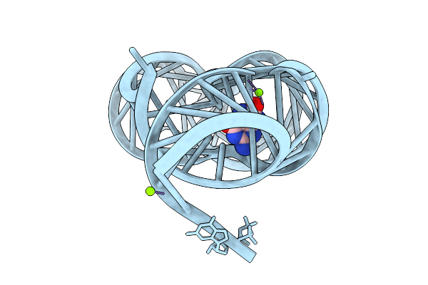
Crystal Structure Of Guanine-Ii Riboswitch In Complex With 7,8-Dihydroneopterin
Organism: Staphylococcus aureus
Method: X-RAY DIFFRACTION Release Date: 2025-11-26 Classification: RNA Ligands: NPR, MG |
 |
Organism: Staphylococcus aureus
Method: X-RAY DIFFRACTION Release Date: 2025-11-26 Classification: RNA Ligands: 7PD, MG |
 |
Organism: Staphylococcus aureus
Method: X-RAY DIFFRACTION Release Date: 2025-11-26 Classification: RNA Ligands: 9QC |
 |
Organism: Staphylococcus aureus
Method: X-RAY DIFFRACTION Release Date: 2025-11-26 Classification: RNA Ligands: ANG, MG |
 |
Organism: Staphylococcus aureus
Method: X-RAY DIFFRACTION Release Date: 2025-11-26 Classification: RNA Ligands: OXG, MG |
 |
Organism: Staphylococcus aureus
Method: X-RAY DIFFRACTION Release Date: 2025-11-26 Classification: RNA Ligands: A1LXN, MG |
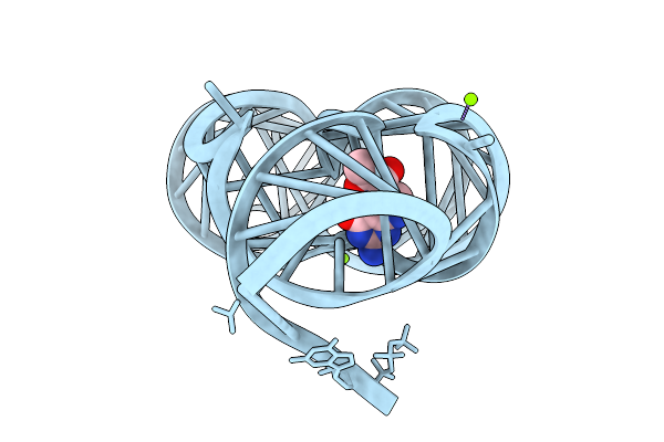 |
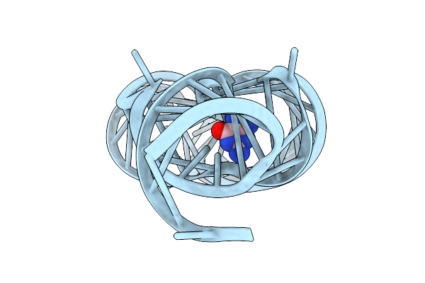
Crystal Structure Of Guanine-Ii Riboswitch In Complex With Tetrahydrobiopterin
Organism: Staphylococcus aureus
Method: X-RAY DIFFRACTION Release Date: 2025-11-26 Classification: RNA Ligands: H4B, MG |