
Search Count: 7
 |
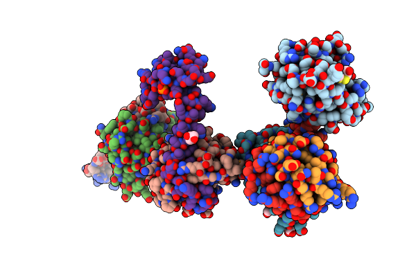
Crystal Structure Of A Mutant Methyl Transferase From Methanosarcina Acetivorans, Northeast Structural Genomics Consortium (Nesg) Target Mvr53-11M
Organism: Methanosarcina acetivorans c2a
Method: X-RAY DIFFRACTION Resolution:2.01 Å Release Date: 2024-01-24 Classification: TRANSFERASE Ligands: SAH |
 |
Organism: Homo sapiens
Method: X-RAY DIFFRACTION Resolution:2.05 Å Release Date: 2024-01-24 Classification: ISOMERASE Ligands: SCN, EDO |
 |
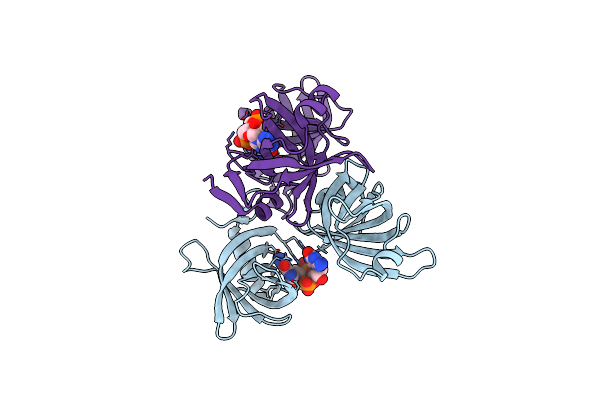
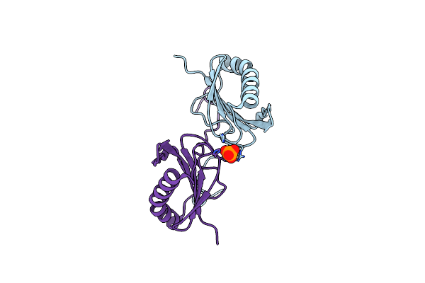
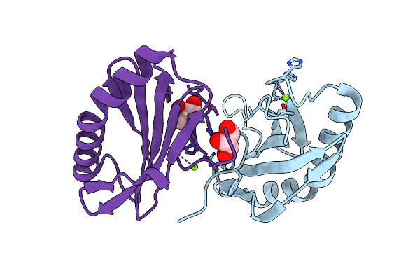
Crystal Structure Of The P1 Bacteriophage Doc Toxin (F68S) In Complex With The Phd Antitoxin (L17M/V39A). Northeast Structural Genomics Targets Er385-Er386
Organism: Bacteriophage p1
Method: X-RAY DIFFRACTION Resolution:2.71 Å Release Date: 2010-08-18 Classification: TOXIN Ligands: CL, HED, GOL, PO4 |
 |
Organism: Vibrio cholerae
Method: X-RAY DIFFRACTION Resolution:1.92 Å Release Date: 2007-10-30 Classification: STRUCTURAL GENOMICS, UNKNOWN FUNCTION Ligands: C2E |
 |
Crystal Structure Of Q83Jn9 From Shigella Flexneri At High Resolution. Northeast Structural Genomics Consortium Target Sfr137.
Organism: Shigella flexneri
Method: X-RAY DIFFRACTION Resolution:1.50 Å Release Date: 2006-10-24 Classification: STRUCTURAL GENOMICS, UNKNOWN FUNCTION Ligands: PO4 |
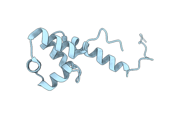 |
Crystal Structure Of The Bacterial Antitoxin Higa From Escherichia Coli At Ph 8.5. Northeast Structural Genomics Target Er390.
Organism: Escherichia coli
Method: X-RAY DIFFRACTION Resolution:1.63 Å Release Date: 2006-09-26 Classification: DNA BINDING PROTEIN |
 |
Crystal Structure Of Yeeu From E. Coli. Northeast Structural Genomics Target Er304
Organism: Escherichia coli
Method: X-RAY DIFFRACTION Resolution:2.10 Å Release Date: 2006-07-18 Classification: STRUCTURAL GENOMICS, UNKNOWN FUNCTION Ligands: CL, MG, GOL |