
Search Count: 12
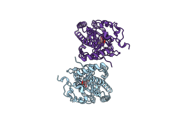 |
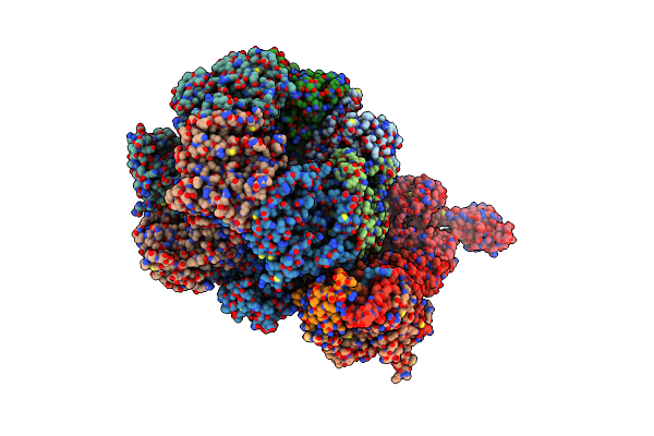
Crystal Structure Of Deoxypodophyllotoxin Synthase From Sinopodophyllum Hexandrum In Complex With 2-Oxoglutarate
Organism: Sinopodophyllum hexandrum
Method: X-RAY DIFFRACTION Resolution:2.09 Å Release Date: 2021-12-15 Classification: BIOSYNTHETIC PROTEIN Ligands: FE, AKG |
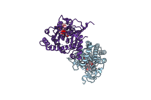 |
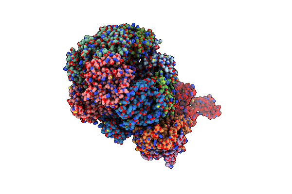
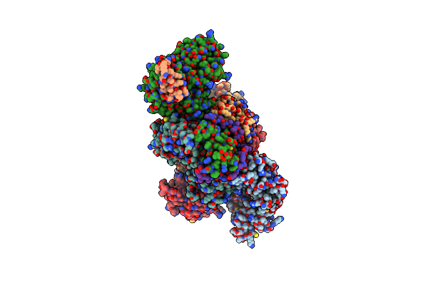
Crystal Structure Of Deoxypodophyllotoxin Synthase From Sinopodophyllum Hexandrum In Complex With Yatein And Succinate
Organism: Sinopodophyllum hexandrum
Method: X-RAY DIFFRACTION Resolution:2.05 Å Release Date: 2021-12-15 Classification: BIOSYNTHETIC PROTEIN Ligands: FE, SIN, YTC, YTN |
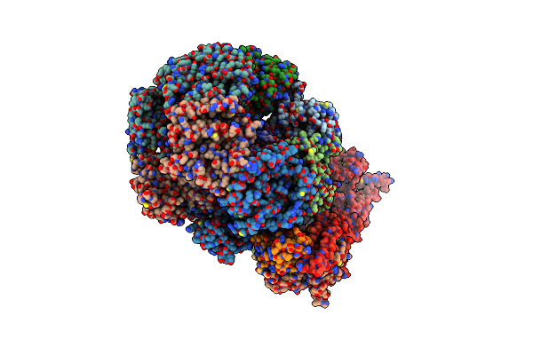 |
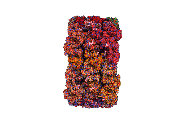
Organism: Drosophila melanogaster
Method: ELECTRON MICROSCOPY Release Date: 2019-09-18 Classification: HYDROLASE Ligands: ADP, ATP |
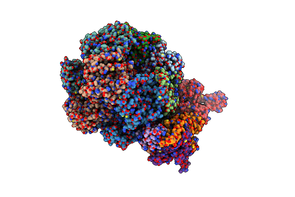 |
Organism: Drosophila melanogaster
Method: ELECTRON MICROSCOPY Release Date: 2019-09-11 Classification: REPLICATION Ligands: ATP, ADP |
 |
Organism: Drosophila melanogaster
Method: ELECTRON MICROSCOPY Release Date: 2019-09-11 Classification: HYDOLASE Ligands: ATP, ADP |
 |
Organism: Drosophila melanogaster
Method: ELECTRON MICROSCOPY Release Date: 2019-09-11 Classification: REPLICATION Ligands: ATP, ADP |
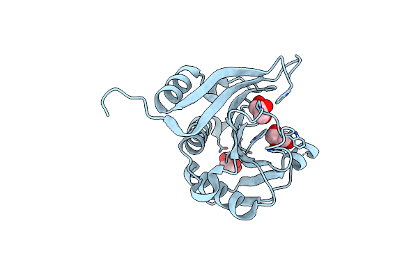 |
Organism: Homo sapiens
Method: X-RAY DIFFRACTION Resolution:1.97 Å Release Date: 2016-10-19 Classification: TRANSLATION Ligands: GOL |
 |
Organism: Todarodes pacificus
Method: X-RAY DIFFRACTION Resolution:3.00 Å Release Date: 2015-10-14 Classification: OXYGEN TRANSPORT Ligands: CUO |
 |
Organism: Drosophila melanogaster
Method: X-RAY DIFFRACTION Resolution:3.50 Å Release Date: 2015-04-01 Classification: DNA BINDING PROTEIN Ligands: K, CL |
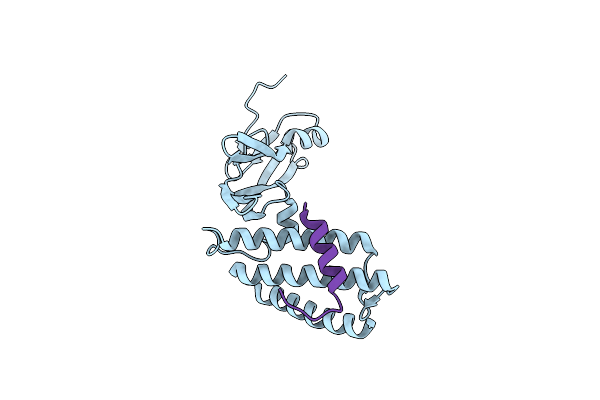 |
Organism: Human papillomavirus type 16, Homo sapiens
Method: X-RAY DIFFRACTION Resolution:1.59 Å Release Date: 2007-01-09 Classification: TRANSCRIPTION |
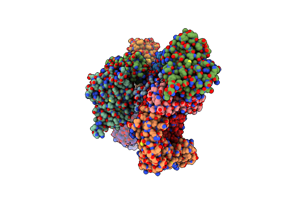 |
The X-Ray Structure Of The Papillomavirus Helicase In Complex With Its Molecular Matchmaker E2
Organism: Human papillomavirus type 18
Method: X-RAY DIFFRACTION Resolution:2.10 Å Release Date: 2004-08-24 Classification: REPLICATION |
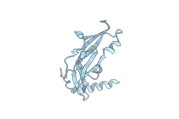 |
2.1 A Crystal Structure Of The Human Papillomavirus Type 18 E2 Activation Domain
Organism: Human papillomavirus type 18
Method: X-RAY DIFFRACTION Resolution:2.10 Å Release Date: 1999-06-21 Classification: VIRAL PROTEIN |