
Search Count: 61
 |
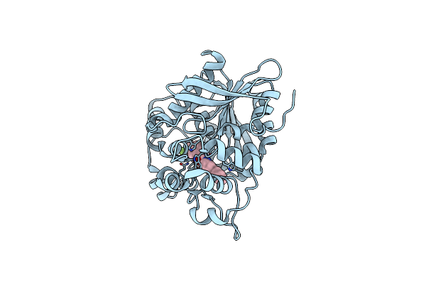
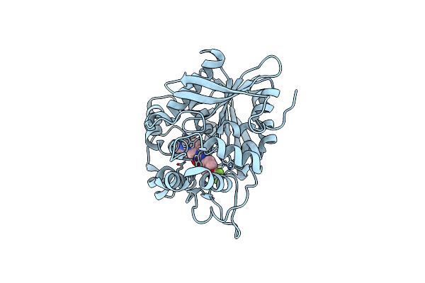
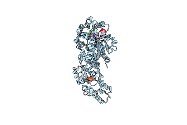
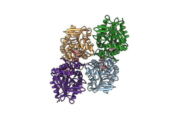
Crystal Structure Of Human Soluble Epoxide Hydrolase C-Terminal Domain In Complex With A Benzohomoadamantane-Based Urea Inhibitor
Organism: Homo sapiens
Method: X-RAY DIFFRACTION Release Date: 2025-04-30 Classification: HYDROLASE Ligands: A1H8Y |
 |
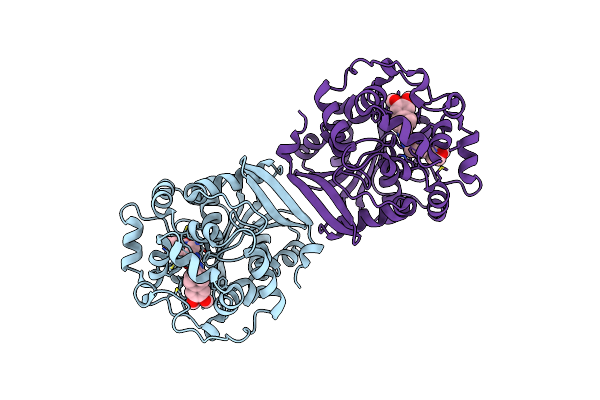
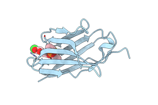
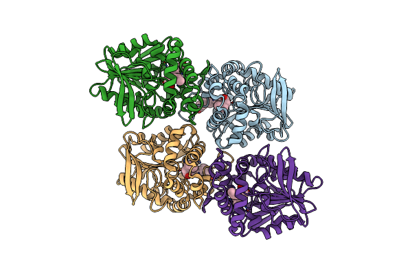
Organism: Camelus bactrianus
Method: X-RAY DIFFRACTION Resolution:2.40 Å Release Date: 2023-10-25 Classification: IMMUNE SYSTEM Ligands: WYN |
 |
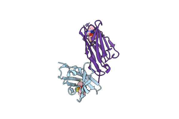
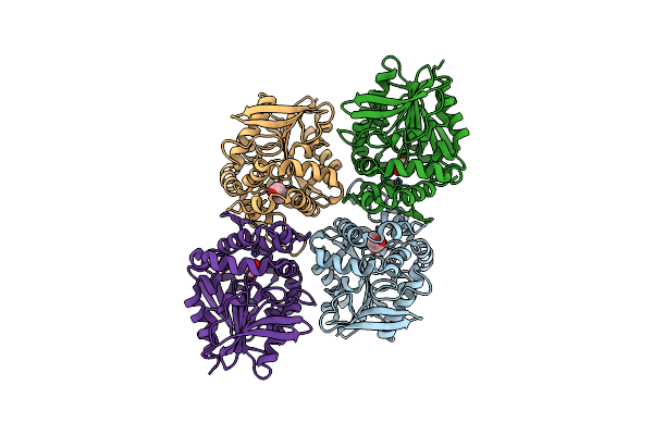
Organism: Camelus bactrianus
Method: X-RAY DIFFRACTION Resolution:1.87 Å Release Date: 2023-10-25 Classification: IMMUNE SYSTEM Ligands: WYS |
 |
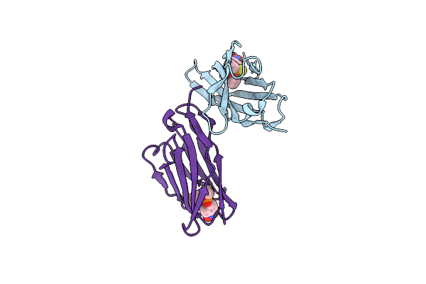
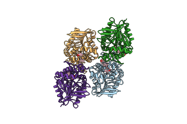
Organism: Camelus bactrianus
Method: X-RAY DIFFRACTION Resolution:1.99 Å Release Date: 2023-10-25 Classification: IMMUNE SYSTEM Ligands: EDO, WYW, NA |
 |
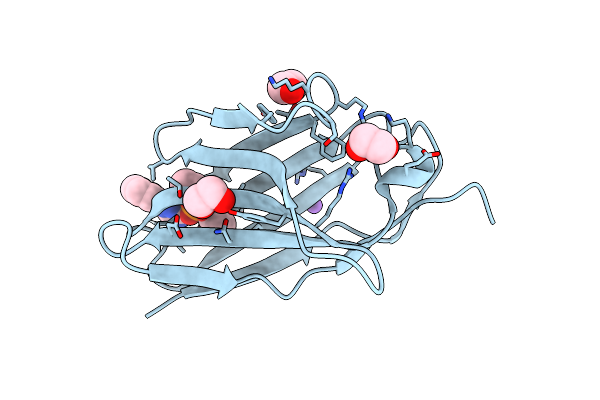
Organism: Camelus bactrianus
Method: X-RAY DIFFRACTION Resolution:2.00 Å Release Date: 2023-10-25 Classification: IMMUNE SYSTEM Ligands: WZ0 |
 |
Organism: Homo sapiens
Method: X-RAY DIFFRACTION Resolution:2.30 Å Release Date: 2022-03-09 Classification: HYDROLASE/HYDROLASE INHIBITOR Ligands: J0U |
 |
Crystal Structure Of An Epoxide Hydrolase From Trichoderma Reesei In Complex With Inhibitor 1
Organism: Trichoderma reesei qm9414
Method: X-RAY DIFFRACTION Resolution:2.20 Å Release Date: 2019-11-20 Classification: HYDROLASE Ligands: K9V |
 |
Crystal Structure Of An Epoxide Hydrolase From Trichoderma Reesei In Complex With Inhibitor 4
Organism: Trichoderma reesei qm9414
Method: X-RAY DIFFRACTION Resolution:2.24 Å Release Date: 2019-11-20 Classification: HYDROLASE Ligands: KC4 |
 |
Crystal Structure Of An Epoxide Hydrolase From Trichoderma Reesei In Complex With Inhibitor 3
Organism: Trichoderma reesei qm9414
Method: X-RAY DIFFRACTION Resolution:1.93 Å Release Date: 2019-11-20 Classification: HYDROLASE/INHIBITOR Ligands: KJ1 |
 |
Crystal Structure Of An Epoxide Hydrolase From Trichoderma Reesei In Complex With Inhibitor 2
Organism: Trichoderma reesei qm9414
Method: X-RAY DIFFRACTION Resolution:2.60 Å Release Date: 2019-11-20 Classification: HYDROLASE/INHIBITOR Ligands: KDS |
 |
Crystal Structure Of An Epoxide Hydrolase From Trichoderma Reesei In Complex With Inhibitor 5
Organism: Trichoderma reesei qm9414
Method: X-RAY DIFFRACTION Resolution:1.72 Å Release Date: 2019-11-20 Classification: HYDROLASE/INHIBITOR Ligands: AUB |
 |
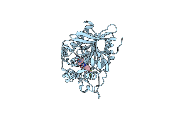
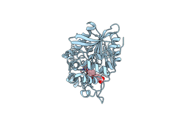
Organism: Homo sapiens
Method: X-RAY DIFFRACTION Resolution:2.00 Å Release Date: 2019-07-03 Classification: HYDROLASE Ligands: MG, G3Q |
 |
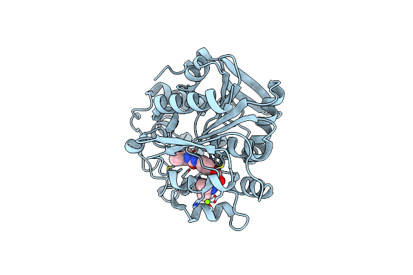
Soluble Epoxide Hydrolase In Complex With 1-(4-((4-(Tert-Butyl)Morpholin-2-Yl)Methoxy)Phenyl)-3-Cyclohexylurea
Organism: Homo sapiens
Method: X-RAY DIFFRACTION Resolution:2.16 Å Release Date: 2019-07-03 Classification: HYDROLASE Ligands: MG, G3T |
 |
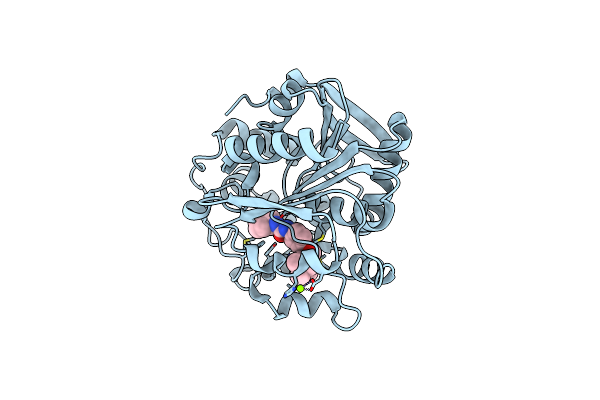
Human Seh Phosphatase In Complex With 3-4-3,4-Dichlorophenyl-5-Phenyl-1,3-Oxazol-2-Yl-Benzoic-Acid
Organism: Homo sapiens
Method: X-RAY DIFFRACTION Resolution:1.55 Å Release Date: 2018-02-28 Classification: HYDROLASE Ligands: MG, 8S9 |
 |
Crystal Structure Of Human Soluble Epoxide Hydrolase Complexed With Trans-4-[4-(3-Trifluoromethoxyphenyl-L-Ureido)-Cyclohexyloxy]-Benzoic Acid.
Organism: Homo sapiens
Method: X-RAY DIFFRACTION Resolution:2.95 Å Release Date: 2018-02-07 Classification: hydrolase/hydrolase inhibitor Ligands: PO4, MG, BXV, CL |
 |
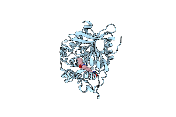
Crystal Structure Of The E153Q Mutant Of The Cftr Inhibitory Factor Cif Containing The Adducted 16,17-Epdpe Hydrolysis Intermediate
Organism: Pseudomonas aeruginosa (strain ucbpp-pa14)
Method: X-RAY DIFFRACTION Resolution:1.80 Å Release Date: 2017-10-18 Classification: HYDROLASE Ligands: 7EZ |
 |
Crystal Structure Of The E153Q Mutant Of The Cftr Inhibitory Factor Cif Containing The Adducted 1,2-Epoxycyclohexane Hydrolysis Intermediate
Organism: Pseudomonas aeruginosa (strain ucbpp-pa14)
Method: X-RAY DIFFRACTION Resolution:1.55 Å Release Date: 2017-10-11 Classification: HYDROLASE Ligands: 40O, 3ZQ |
 |
Crystal Structure Of The E153Q Mutant Of The Cftr Inhibitory Factor Cif Containing The Adducted Cis-Stilbene Oxide Hydrolysis Intermediate
Organism: Pseudomonas aeruginosa (strain ucbpp-pa14)
Method: X-RAY DIFFRACTION Resolution:1.75 Å Release Date: 2017-10-11 Classification: HYDROLASE Ligands: 7F6 |
 |
Crystal Structure Of The E153Q Mutant Of The Cftr Inhibitory Factor Cif Containing The Adducted 19,20-Epdpe Hydrolysis Intermediate
Organism: Pseudomonas aeruginosa (strain ucbpp-pa14)
Method: X-RAY DIFFRACTION Resolution:1.75 Å Release Date: 2017-10-11 Classification: hydrolase/hydrolase inhibitor Ligands: 7HW |
 |
Crystal Structure Of The E153Q Mutant Of The Cftr Inhibitory Factor Cif Containing The Adducted 14,15-Epete Hydrolysis Intermediate
Organism: Pseudomonas aeruginosa (strain ucbpp-pa14)
Method: X-RAY DIFFRACTION Resolution:1.75 Å Release Date: 2017-10-11 Classification: HYDROLASE Ligands: 7LE |