
Search Count: 33
 |
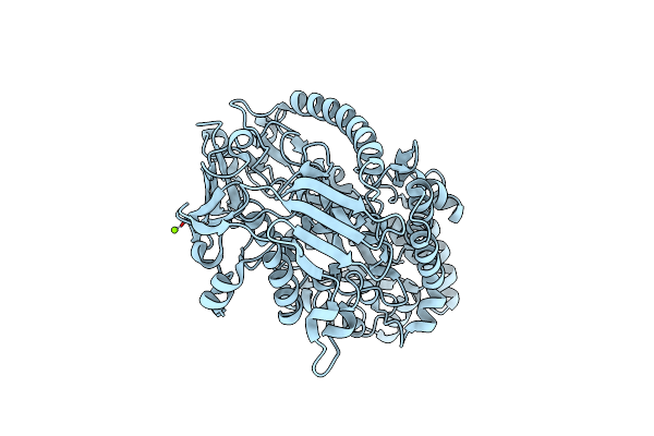
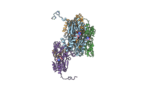
Organism: Streptococcus canis
Method: X-RAY DIFFRACTION Resolution:2.70 Å Release Date: 2025-10-01 Classification: IMMUNE SYSTEM Ligands: SO4 |
 |
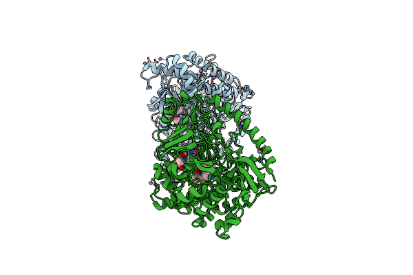
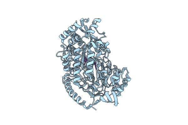
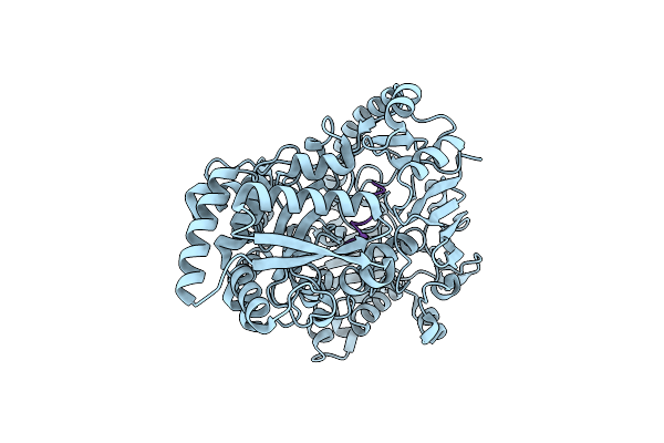
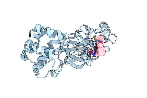
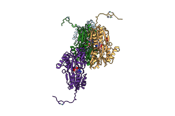
Structure Of The Truncated Version Of Idec Protease C94S From Streptococcus Canis
Organism: Streptococcus canis
Method: X-RAY DIFFRACTION Resolution:2.25 Å Release Date: 2025-10-01 Classification: IMMUNE SYSTEM |
 |
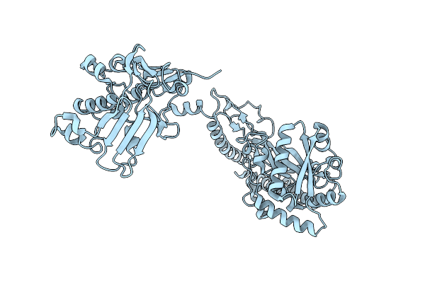
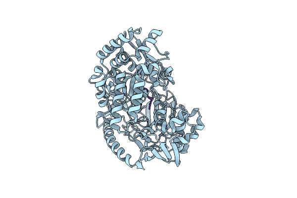
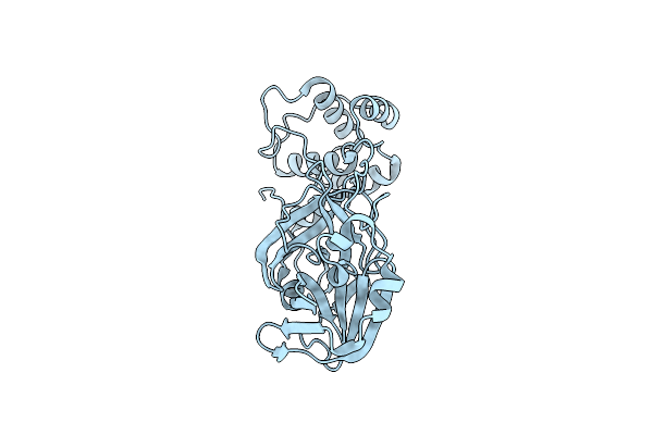
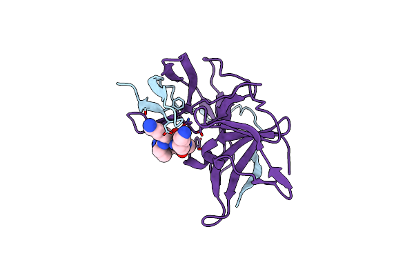
Crystal Structure Of The Pneumococcal Substrate-Binding Protein Alid In Open Conformation
Organism: Streptococcus pneumoniae
Method: X-RAY DIFFRACTION Resolution:1.80 Å Release Date: 2024-05-22 Classification: PEPTIDE BINDING PROTEIN Ligands: MG |
 |
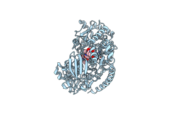
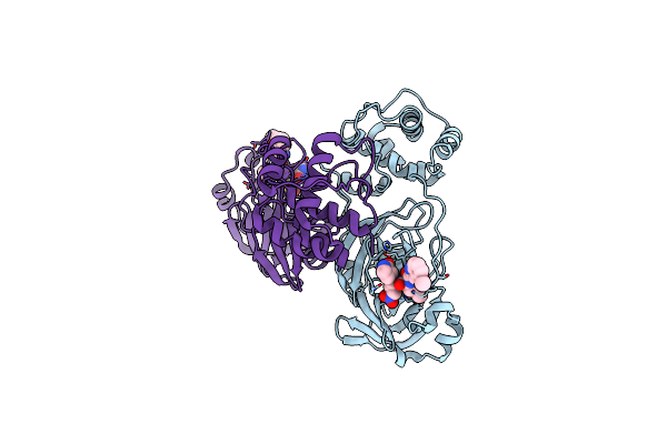
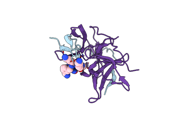
Crystal Structure Of The Pneumococcal Substrate-Binding Protein Alid In Closed Conformation In Complex With Peptide 1
Organism: Streptococcus pneumoniae, Prevotella
Method: X-RAY DIFFRACTION Resolution:2.10 Å Release Date: 2024-05-22 Classification: PEPTIDE BINDING PROTEIN Ligands: ZN |
 |
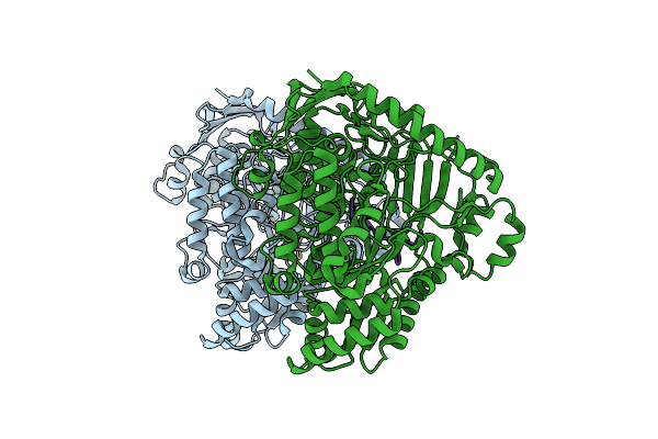
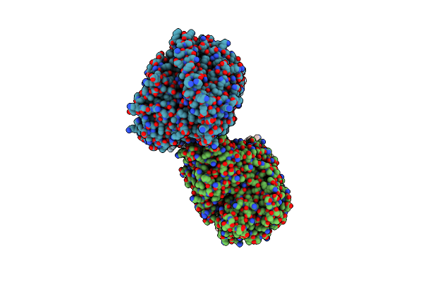
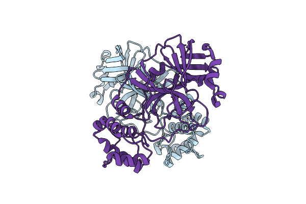
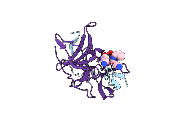
Crystal Structure Of The Pneumococcal Substrate-Binding Protein Alic As A Domain-Swapped Dimer
Organism: Streptococcus pneumoniae
Method: X-RAY DIFFRACTION Resolution:2.38 Å Release Date: 2024-05-22 Classification: PEPTIDE BINDING PROTEIN |
 |
Crystal Structure Of The Pneumococcal Substrate-Binding Protein Alib In Complex With An Unknown Peptide
Organism: Streptococcus pneumoniae, Escherichia coli bl21(de3)
Method: X-RAY DIFFRACTION Resolution:1.65 Å Release Date: 2024-05-22 Classification: PEPTIDE BINDING PROTEIN |
 |
Crystal Structure Of The Pneumococcal Substrate-Binding Protein Alib In Complex With Peptide 2
Organism: Streptococcus pneumoniae, Synthetic construct
Method: X-RAY DIFFRACTION Resolution:2.29 Å Release Date: 2024-05-22 Classification: PEPTIDE BINDING PROTEIN |
 |
Crystal Structure Of The Pneumococcal Substrate-Binding Protein Alib In Complex With Peptide 3
Organism: Streptococcus pneumoniae, Synthetic construct
Method: X-RAY DIFFRACTION Resolution:1.66 Å Release Date: 2024-05-22 Classification: PEPTIDE BINDING PROTEIN |
 |
Crystal Structure Of The Pneumococcal Substrate-Binding Protein Alib In Complex With Peptide 4
Organism: Streptococcus pneumoniae, Synthetic construct
Method: X-RAY DIFFRACTION Resolution:1.49 Å Release Date: 2024-05-22 Classification: PEPTIDE BINDING PROTEIN |
 |
Crystal Structure Of The Pneumococcal Substrate-Binding Protein Amia In Complex With Peptide 5
Organism: Streptococcus pneumoniae, Bacteria
Method: X-RAY DIFFRACTION Resolution:1.76 Å Release Date: 2024-05-22 Classification: PEPTIDE BINDING PROTEIN |
 |
Crystal Structure Of The Pneumococcal Substrate-Binding Protein Amia In Complex With An Unknown Peptide
Organism: Streptococcus pneumoniae, Escherichia coli
Method: X-RAY DIFFRACTION Resolution:1.50 Å Release Date: 2023-12-20 Classification: PEPTIDE BINDING PROTEIN |
 |
Organism: Severe acute respiratory syndrome coronavirus 2
Method: X-RAY DIFFRACTION Resolution:1.99 Å Release Date: 2023-11-08 Classification: VIRAL PROTEIN |
 |
Organism: Severe acute respiratory syndrome coronavirus 2
Method: X-RAY DIFFRACTION Resolution:1.86 Å Release Date: 2023-11-08 Classification: VIRAL PROTEIN |
 |
Organism: Severe acute respiratory syndrome coronavirus 2
Method: X-RAY DIFFRACTION Resolution:1.98 Å Release Date: 2023-11-08 Classification: VIRAL PROTEIN Ligands: QQL |
 |
Organism: Severe acute respiratory syndrome coronavirus 2
Method: X-RAY DIFFRACTION Resolution:1.62 Å Release Date: 2023-11-08 Classification: VIRAL PROTEIN Ligands: QH0 |
 |
Crystal Structure Of Unlinked Ns2B_Ns3 Protease From Zika Virus In Complex With Inhibitor Mi-2224
Organism: Zika virus
Method: X-RAY DIFFRACTION Resolution:1.75 Å Release Date: 2022-12-28 Classification: VIRAL PROTEIN Ligands: IXU |
 |
Crystal Structure Of Unlinked Ns2B_Ns3 Protease From Zika Virus In Complex With Inhibitor Mi-2223
Organism: Zika virus
Method: X-RAY DIFFRACTION Resolution:1.61 Å Release Date: 2022-12-28 Classification: VIRAL PROTEIN Ligands: IY3 |
 |
Crystal Structure Of Unlinked Ns2B_Ns3 Protease From Zika Virus In Complex With Inhibitor Mi-2113
Organism: Zika virus
Method: X-RAY DIFFRACTION Resolution:2.15 Å Release Date: 2022-12-28 Classification: VIRAL PROTEIN Ligands: IXJ |
 |
Organism: Streptococcus pneumoniae
Method: X-RAY DIFFRACTION Resolution:2.50 Å Release Date: 2021-02-10 Classification: TRANSPORT PROTEIN Ligands: NI, ADN, ACT |
 |
Organism: Streptococcus pneumoniae
Method: X-RAY DIFFRACTION Resolution:2.28 Å Release Date: 2021-02-10 Classification: TRANSPORT PROTEIN Ligands: GMP, NI |