
Search Count: 131
 |
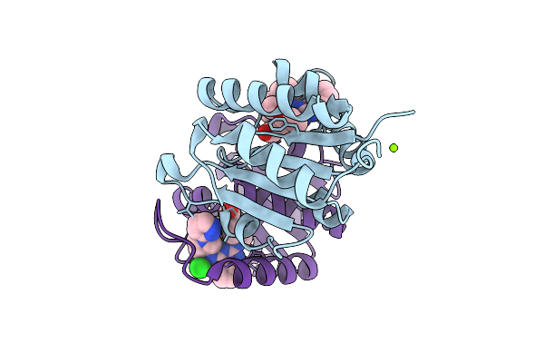
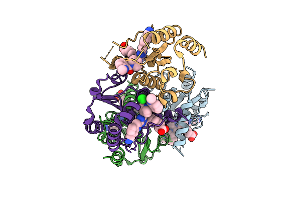
Organism: Homo sapiens
Method: X-RAY DIFFRACTION Resolution:2.02 Å Release Date: 2025-12-24 Classification: HYDROLASE Ligands: A1JH5 |
 |
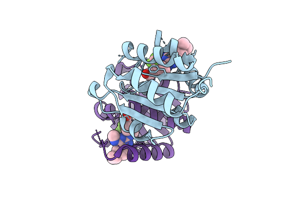
Organism: Homo sapiens
Method: X-RAY DIFFRACTION Resolution:1.51 Å Release Date: 2025-12-24 Classification: HYDROLASE Ligands: A1JKT, EDO |
 |
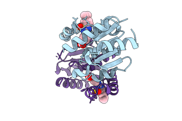
Organism: Homo sapiens
Method: X-RAY DIFFRACTION Resolution:1.50 Å Release Date: 2025-12-24 Classification: HYDROLASE Ligands: A1JKZ |
 |
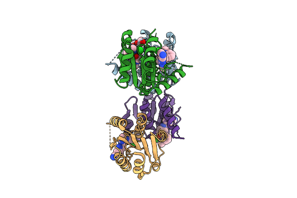
Organism: Homo sapiens
Method: X-RAY DIFFRACTION Resolution:1.77 Å Release Date: 2025-12-24 Classification: HYDROLASE Ligands: CL, A1JK6 |
 |
Organism: Mycolicibacterium fortuitum
Method: X-RAY DIFFRACTION Resolution:1.32 Å Release Date: 2025-12-17 Classification: FLAVOPROTEIN |
 |
Organism: Mycolicibacterium fortuitum
Method: X-RAY DIFFRACTION Resolution:1.42 Å Release Date: 2025-12-17 Classification: FLAVOPROTEIN |
 |
Organism: Homo sapiens
Method: X-RAY DIFFRACTION Resolution:1.60 Å Release Date: 2025-11-12 Classification: HYDROLASE Ligands: A1IZR |
 |
Organism: Homo sapiens
Method: X-RAY DIFFRACTION Resolution:1.22 Å Release Date: 2025-11-12 Classification: HYDROLASE Ligands: MG, A1IZW, CL |
 |
Organism: Homo sapiens
Method: X-RAY DIFFRACTION Resolution:1.21 Å Release Date: 2025-11-12 Classification: HYDROLASE Ligands: A1IZX |
 |
Organism: Homo sapiens
Method: X-RAY DIFFRACTION Resolution:1.49 Å Release Date: 2025-11-12 Classification: HYDROLASE Ligands: A1I0K |
 |
Organism: Homo sapiens
Method: X-RAY DIFFRACTION Resolution:1.72 Å Release Date: 2025-11-12 Classification: HYDROLASE Ligands: EOH, EDO, A1I1M, CL |
 |
Organism: Homo sapiens
Method: X-RAY DIFFRACTION Resolution:2.21 Å Release Date: 2025-11-12 Classification: HYDROLASE Ligands: CL, A1I1U |
 |
Mouse Otoferlin (216-1931) In Complex With An Msp2N2 Lipid Nanodisc (30 Mol% Dops, 10 Mol% Pi(4,5)P2)
Organism: Mus musculus
Method: ELECTRON MICROSCOPY Release Date: 2025-11-12 Classification: MEMBRANE PROTEIN Ligands: CA, PSF |
 |
Organism: Homo sapiens
Method: X-RAY DIFFRACTION Resolution:2.07 Å Release Date: 2025-11-12 Classification: HYDROLASE Ligands: A1JFX |
 |
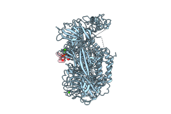
Mouse Otoferlin (216-1931) In The Lipid-Free, Ca2+-Bound State, "Open" Conformation (Class 2)
Organism: Mus musculus
Method: ELECTRON MICROSCOPY Release Date: 2025-11-05 Classification: MEMBRANE PROTEIN Ligands: CA |
 |
Mouse Otoferlin (216-1931) In Complex With A Lipid Nanodisc (Comprising 25% Ps And 5% Pip2)
Organism: Mus musculus
Method: ELECTRON MICROSCOPY Release Date: 2025-11-05 Classification: MEMBRANE PROTEIN Ligands: CA, PSF |
 |
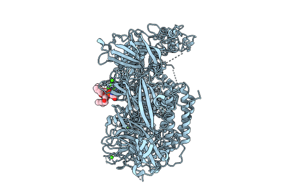
Mouse Otoferlin (216-1931) In The Lipid-Free Ca2+-Bound State, "Open" Conformation (Class 1)
Organism: Mus musculus
Method: ELECTRON MICROSCOPY Release Date: 2025-11-05 Classification: MEMBRANE PROTEIN Ligands: CA |
 |
Mouse Otoferlin (Residues 216-1931) In The Lipid-Bound State (Merged Datasets)
Organism: Mus musculus
Method: ELECTRON MICROSCOPY Release Date: 2025-11-05 Classification: MEMBRANE PROTEIN Ligands: CA, PSF |
 |
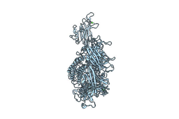
Mouse Otoferlin (216-1931) In The Lipid-Free, Ca2+-Free State ("Loose" Conformation)
Organism: Mus musculus
Method: ELECTRON MICROSCOPY Release Date: 2025-11-05 Classification: MEMBRANE PROTEIN |
 |
Mouse Otoferlin (216-1931) In The Lipid-Free Ca2+-Bound State, "Closed-Like" Conformation
Organism: Mus musculus
Method: ELECTRON MICROSCOPY Release Date: 2025-11-05 Classification: MEMBRANE PROTEIN Ligands: CA |