Search Count: 13
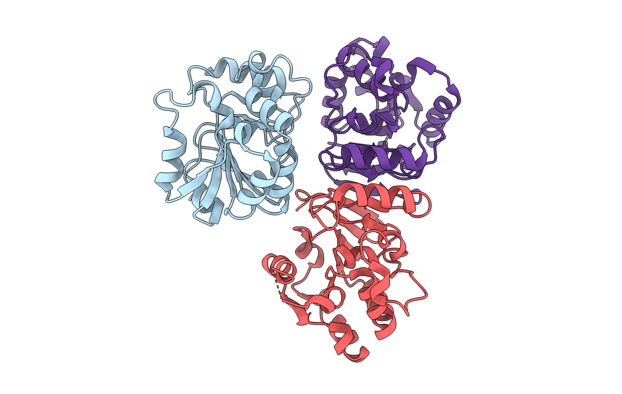 |
Crystal Structure Of Peptidase E From Deinococcus Radiodurans In P6422 Space Group
Organism: Deinococcus radiodurans
Method: X-RAY DIFFRACTION Resolution:2.70 Å Release Date: 2019-11-20 Classification: HYDROLASE |
 |
Organism: Deinococcus radiodurans r1
Method: X-RAY DIFFRACTION Resolution:2.00 Å Release Date: 2019-06-26 Classification: HYDROLASE |
 |
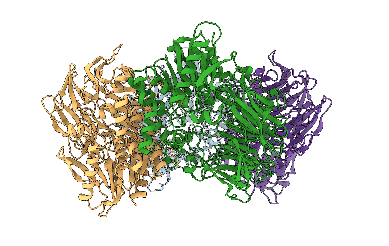
Crystal Structure Of S9 Peptidase (Inactive Form) From Deinococcus Radiodurans R1
Organism: Deinococcus radiodurans (strain atcc 13939 / dsm 20539 / jcm 16871 / lmg 4051 / nbrc 15346 / ncimb 9279 / r1 / vkm b-1422)
Method: X-RAY DIFFRACTION Resolution:2.30 Å Release Date: 2018-11-14 Classification: HYDROLASE Ligands: ACT |
 |
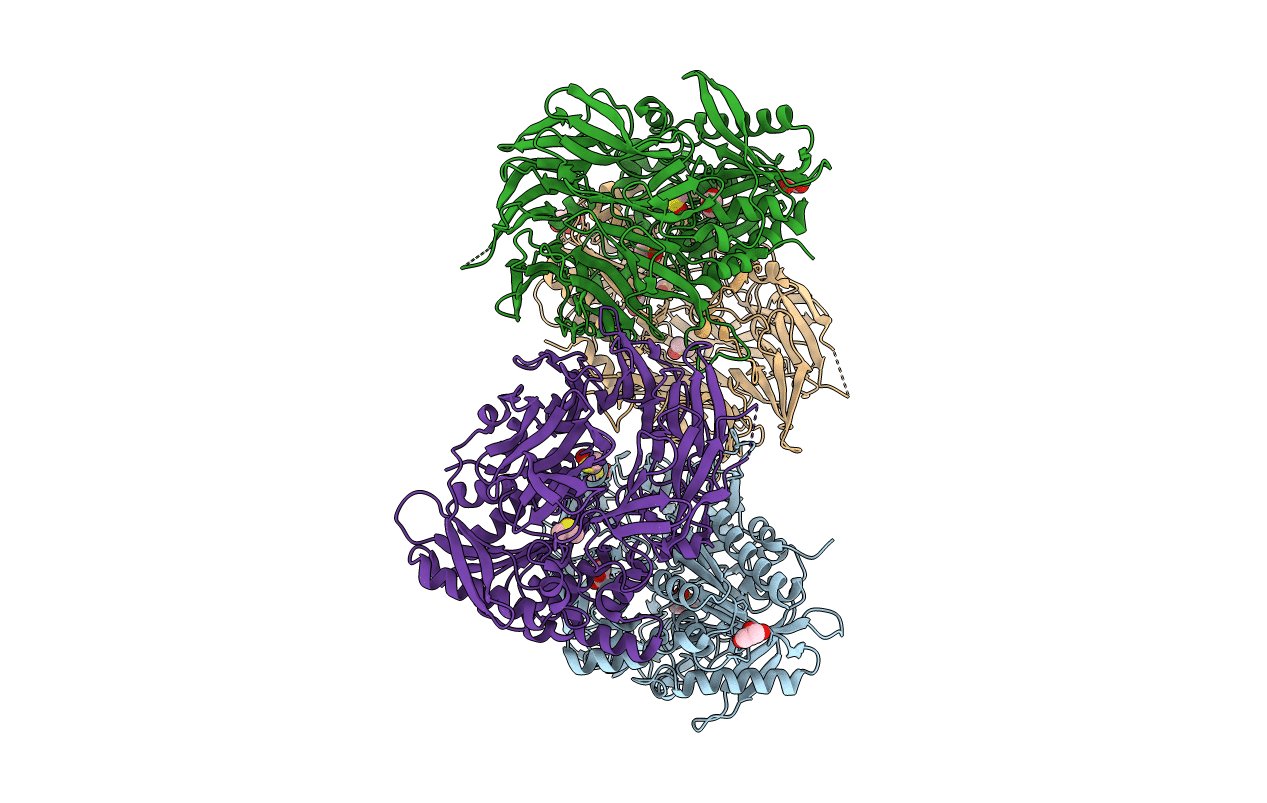
Crystal Structure Of S9 Peptidase (Active Form) From Deinococcus Radiodurans R1
Organism: Deinococcus radiodurans (strain atcc 13939 / dsm 20539 / jcm 16871 / lmg 4051 / nbrc 15346 / ncimb 9279 / r1 / vkm b-1422)
Method: X-RAY DIFFRACTION Resolution:2.30 Å Release Date: 2018-11-14 Classification: HYDROLASE |
 |
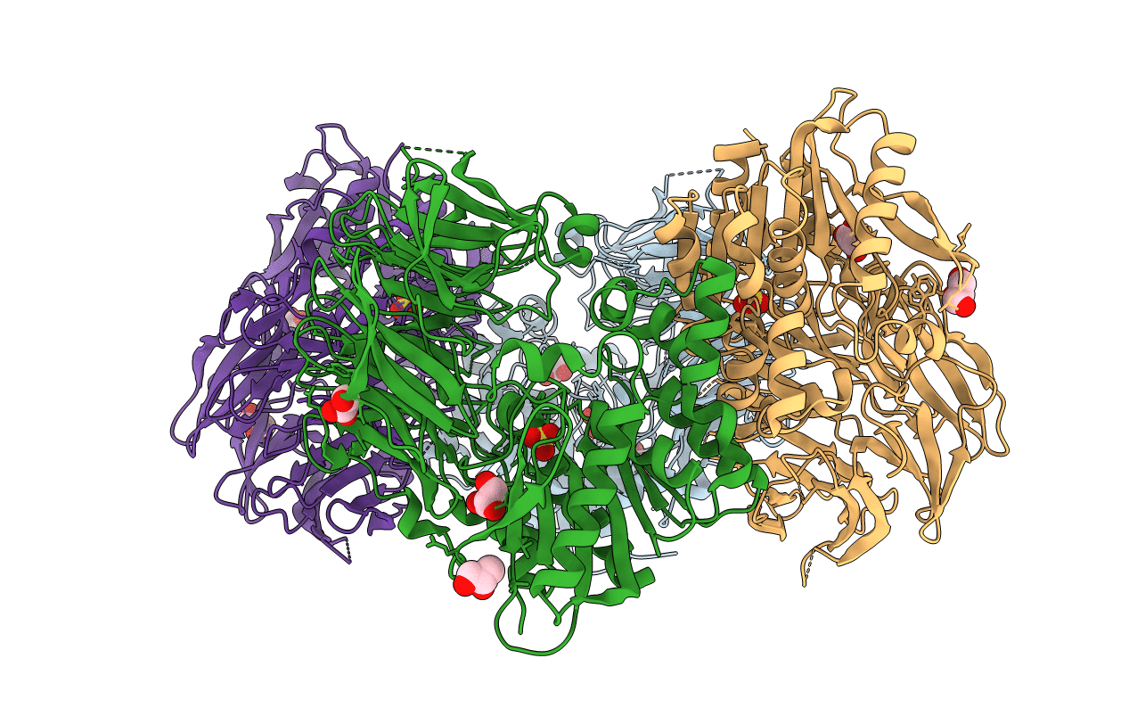
Crystal Structure Of S9 Peptidase Mutant (S514A) From Deinococcus Radiodurans R1
Organism: Deinococcus radiodurans (strain atcc 13939 / dsm 20539 / jcm 16871 / lmg 4051 / nbrc 15346 / ncimb 9279 / r1 / vkm b-1422)
Method: X-RAY DIFFRACTION Resolution:1.70 Å Release Date: 2018-11-14 Classification: HYDROLASE Ligands: DMS, GOL |
 |
Crystal Structure Of S9 Peptidase (Inactive State)From Deinococcus Radiodurans R1 In P212121
Organism: Deinococcus radiodurans str. r1
Method: X-RAY DIFFRACTION Resolution:2.40 Å Release Date: 2018-11-14 Classification: HYDROLASE Ligands: GOL |
 |
Crystal Structure Of Inactive State Of S9 Peptidase From Deinococcus Radiodurans R1 (Pmsf Treated)
Organism: Deinococcus radiodurans str. r1
Method: X-RAY DIFFRACTION Resolution:2.30 Å Release Date: 2018-11-14 Classification: HYDROLASE Ligands: GOL, SO4 |
 |
Crystal Structure Of S9 Peptidase (S514A Mutant In Inactive State) From Deinococcus Radiodurans R1
Organism: Deinococcus radiodurans str. r1
Method: X-RAY DIFFRACTION Resolution:2.60 Å Release Date: 2018-11-14 Classification: HYDROLASE Ligands: GOL |
 |
Crystal Structure Of Substrate-Bound S9 Peptidase (S514A Mutant) From Deinococcus Radiodurans
Organism: Deinococcus radiodurans r1, Deinococcus radiodurans
Method: X-RAY DIFFRACTION Resolution:2.30 Å Release Date: 2018-11-14 Classification: HYDROLASE Ligands: GOL |
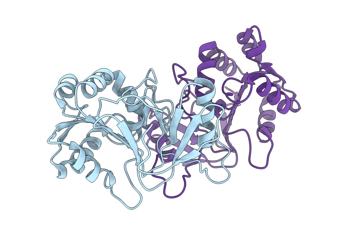 |
Crystal Structure Of Peptidase E With Ordered Active Site Loop From Salmonella Enterica
Organism: Salmonella typhimurium (strain lt2 / sgsc1412 / atcc 700720)
Method: X-RAY DIFFRACTION Resolution:1.90 Å Release Date: 2018-10-31 Classification: HYDROLASE |
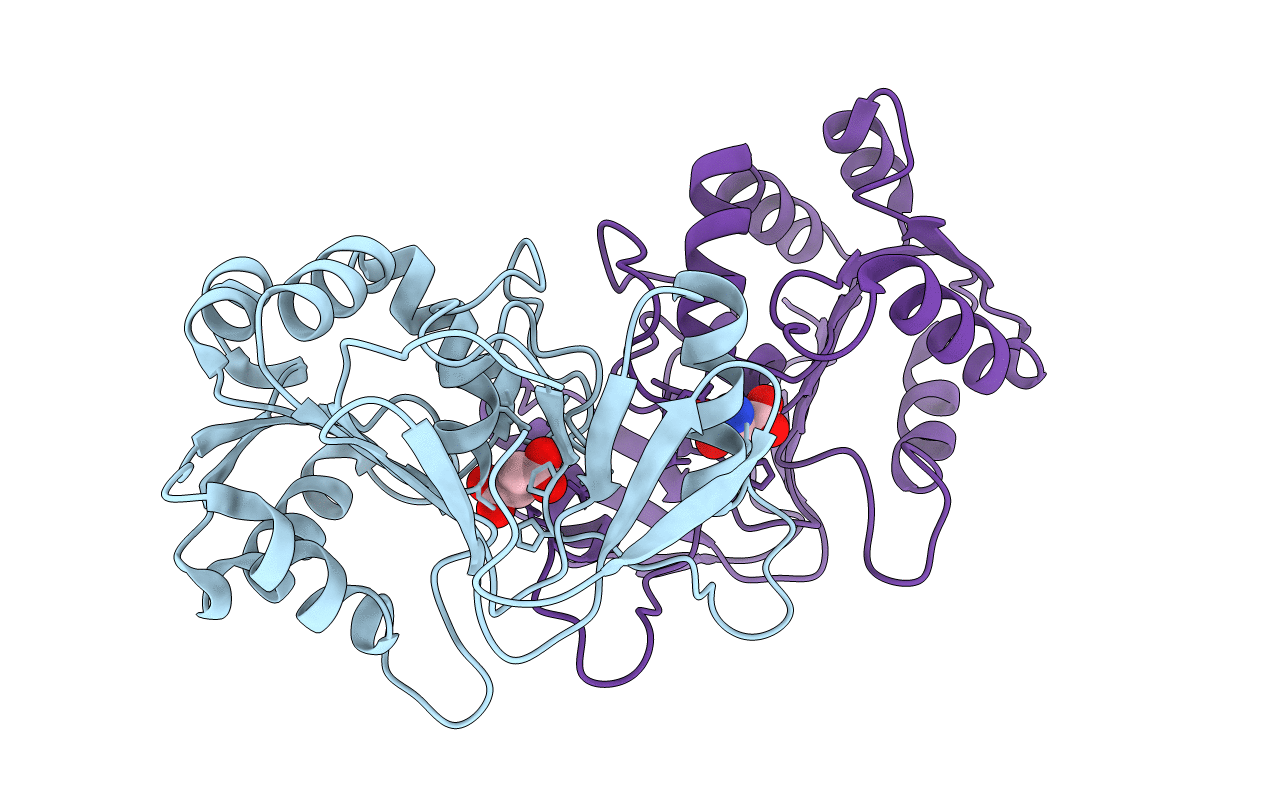 |
Organism: Salmonella typhimurium (strain lt2 / sgsc1412 / atcc 700720)
Method: X-RAY DIFFRACTION Resolution:1.83 Å Release Date: 2018-10-24 Classification: HYDROLASE Ligands: ASP |
 |
Organism: Homo sapiens
Method: X-RAY DIFFRACTION Resolution:2.30 Å Release Date: 2014-07-30 Classification: DNA BINDING PROTEIN Ligands: EDO |
 |
Organism: Homo sapiens
Method: X-RAY DIFFRACTION Resolution:2.25 Å Release Date: 2014-07-30 Classification: DNA BINDING PROTEIN |

