
Search Count: 41
 |
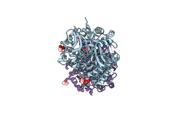
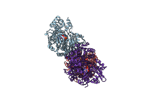
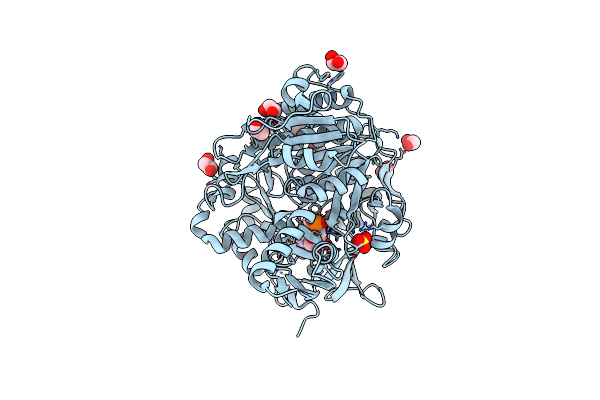
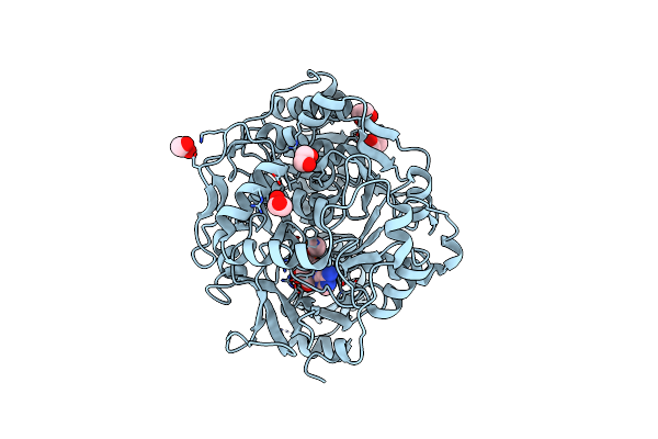
Crystal Structure Of Acetyl-Coa Synthetase From Cryptococcus Neoformans H99 In Complex With Inhibitor Hgn-1310 (Dd3-027)
Organism: Cryptococcus neoformans var. grubii
Method: X-RAY DIFFRACTION Release Date: 2025-10-29 Classification: LIGASE Ligands: GOL, SO4, CL, A1AV1 |
 |
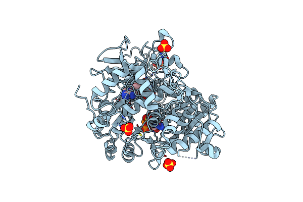
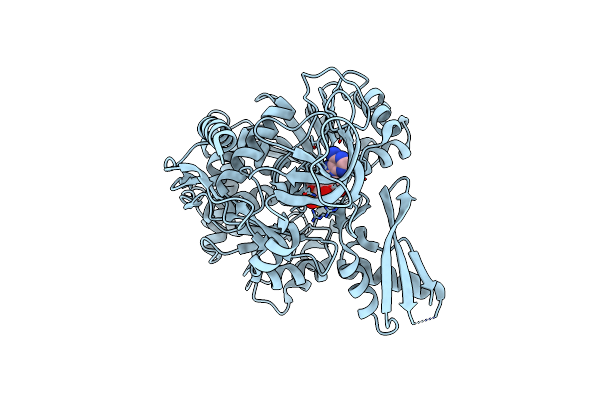
Crystal Structure Of Acetyl-Coa Synthetase From Cryptococcus Neoformans H99 In Complex With An Ethylsulfamide Amp Inhibitor
Organism: Cryptococcus neoformans var. grubii
Method: X-RAY DIFFRACTION Release Date: 2023-12-13 Classification: LIGASE Ligands: YDO, CL |
 |
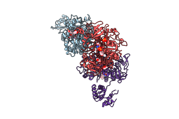
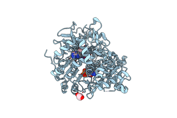
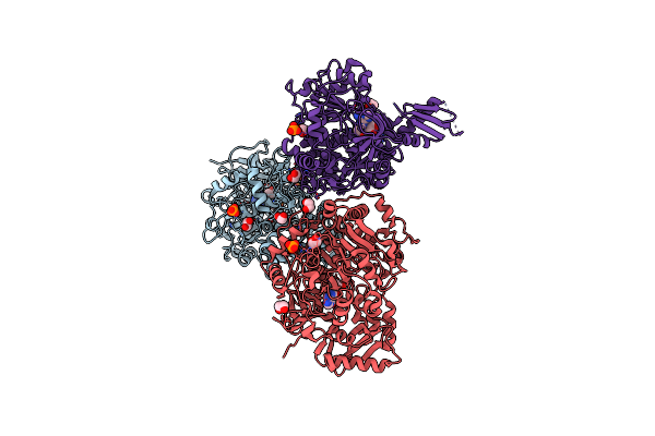
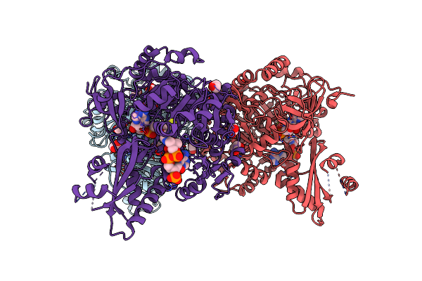
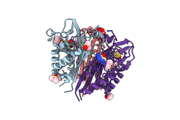
Crystal Structure Of Acetyl-Coa Synthetase 2 In Complex With Amp And Coa From Candida Albicans
Organism: Candida albicans
Method: X-RAY DIFFRACTION Resolution:2.70 Å Release Date: 2023-12-06 Classification: LIGASE Ligands: AMP, COA, ACY, GOL |
 |
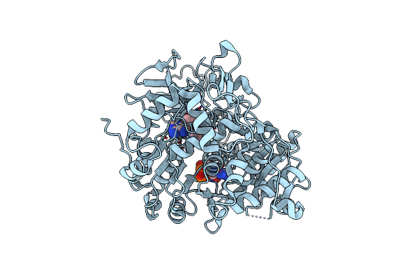
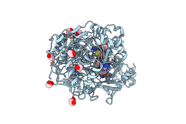
Crystal Structure Of Acetyl-Coenzyme A Synthetase From Leishmania Infantum (Ethyl Amp Bound)
Organism: Leishmania infantum
Method: X-RAY DIFFRACTION Resolution:1.55 Å Release Date: 2023-09-13 Classification: LIGASE Ligands: PEG, WTA |
 |
Crystal Structure Of Acetyl-Coenzyme A Synthetase From Leishmania Infantum (Ethyl Amp Bound, P21 Form)
Organism: Leishmania infantum
Method: X-RAY DIFFRACTION Resolution:2.52 Å Release Date: 2023-09-13 Classification: LIGASE Ligands: CAC, GOL, CL, WTA |
 |
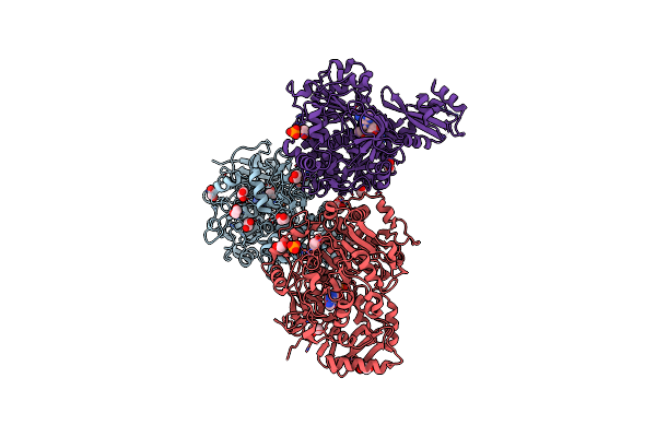
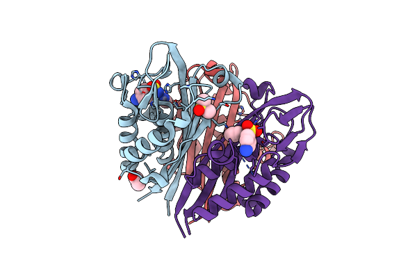
Crystal Structure Of Acetyl-Coenzyme A Synthetase From Leishmania Infantum (Coa And Amp Bound)
Organism: Leishmania infantum
Method: X-RAY DIFFRACTION Resolution:1.65 Å Release Date: 2023-09-13 Classification: LIGASE Ligands: SO4, CL, AMP, COA |
 |
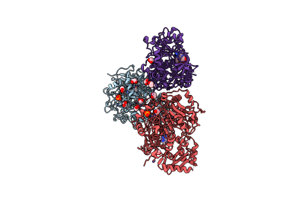
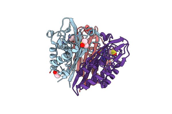
Crystal Structure Of Acetyl-Coenzyme A Synthetase From Leishmania Infantum (Coa, Amp And Potassium Bound)
Organism: Leishmania infantum
Method: X-RAY DIFFRACTION Resolution:1.97 Å Release Date: 2023-09-13 Classification: LIGASE Ligands: K, CL, GOL, AMP, COA |
 |
Crystal Structure Of Acetyl-Coenzyme A Synthetase From Leishmania Infantum (Amp, Acetate And Coa Bound)
Organism: Leishmania infantum
Method: X-RAY DIFFRACTION Resolution:1.70 Å Release Date: 2023-04-19 Classification: LIGASE Ligands: AMP, ACT, COA |
 |
Crystal Structure Of Acetyl-Coa Synthetase In Complex With A Cyclopropyl Ester Amp Inhibitor From Cryptococcus Neoformans H99
Organism: Cryptococcus neoformans
Method: X-RAY DIFFRACTION Release Date: 2023-02-15 Classification: LIGASE Ligands: EDO, YHK |
 |
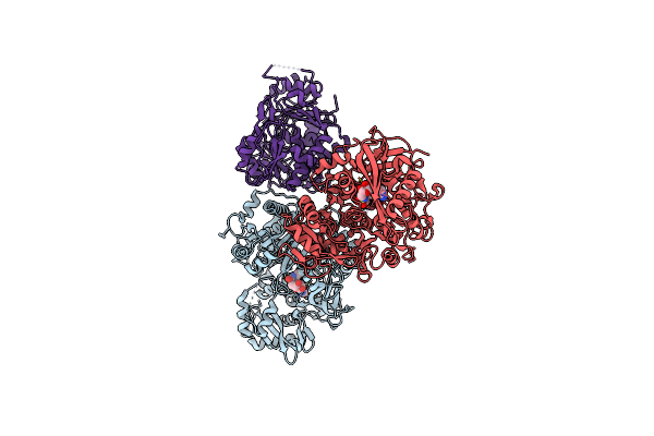
Crystal Structure Of Acetyl-Coenzyme A Synthetase From Legionella Pneumophila Philadelphia 1 In Complex With Ethyl-Amp
Organism: Legionella pneumophila subsp. pneumophila (strain philadelphia 1 / atcc 33152 / dsm 7513)
Method: X-RAY DIFFRACTION Resolution:2.40 Å Release Date: 2021-05-26 Classification: LIGASE Ligands: WTA, EDO |
 |
Crystal Structure Of Acetyl-Coa Synthetase In Complex With Adenosine-5'-Ethylphosphate From Cryptococcus Neoformans Var. Grubii Serotype A (H99)
Organism: Cryptococcus neoformans var. grubii serotype a (strain h99 / atcc 208821 / cbs 10515 / fgsc 9487)
Method: X-RAY DIFFRACTION Resolution:1.80 Å Release Date: 2020-12-30 Classification: LIGASE Ligands: WTA, EDO, PO4 |
 |
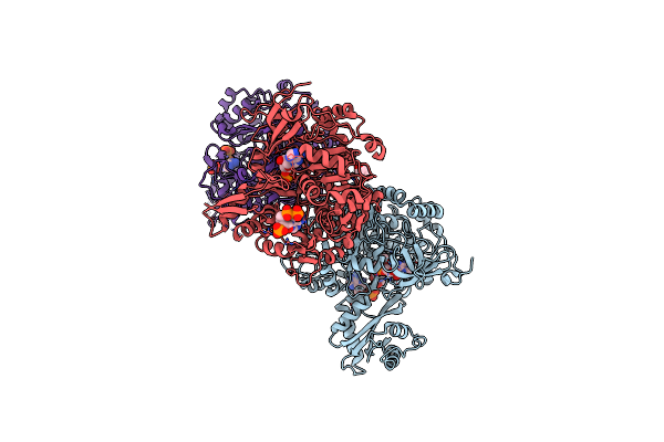
Crystal Structure Of Acetyl-Coa Synthetase In Complex With Adenosine-5'-Ethylphosphate And Co-Enzyme A From Coccidioides Immitis Rs
Organism: Coccidioides immitis (strain rs)
Method: X-RAY DIFFRACTION Resolution:1.90 Å Release Date: 2020-12-30 Classification: LIGASE Ligands: COA, WTA, EDO |
 |
Crystal Structure Of Acetyl-Coa Synthetase In Complex With Adenosine-5'-Ethylphosphate And Co-Enzyme A From Coccidioides Immitis Rs
Organism: Coccidioides immitis (strain rs)
Method: X-RAY DIFFRACTION Resolution:2.10 Å Release Date: 2020-12-30 Classification: LIGASE Ligands: WTA, EDO, SO4 |
 |
Crystal Structure Of Acetyl-Coa Synthetase In Complex With Adenosine-5'-Methylphosphate And Co-Enzyme A From Coccidioides Immitis Rs
Organism: Coccidioides immitis (strain rs)
Method: X-RAY DIFFRACTION Resolution:2.15 Å Release Date: 2020-12-30 Classification: LIGASE Ligands: EDO, COA, XM7 |
 |
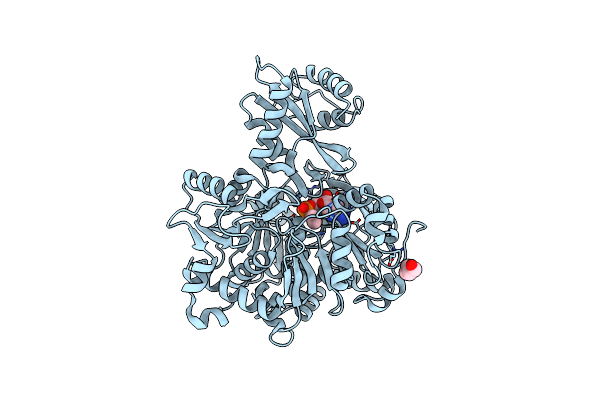
Crystal Structure Of Acetyl-Coa Synthetase In Complex With Acetyl Adenylate From Cryptococcus Neoformans H99
Organism: Cryptococcus neoformans var. grubii serotype a (strain h99 / atcc 208821 / cbs 10515 / fgsc 9487)
Method: X-RAY DIFFRACTION Resolution:2.20 Å Release Date: 2020-12-30 Classification: LIGASE Ligands: 6R9, PO4, EDO, K |
 |
Crystal Structure Of Acetyl-Coa Synthetase In Complex With Adenosine-5'-Butylphosphate From Cryptococcus Neoformans Var. Grubii Serotype A (H99)
Organism: Cryptococcus neoformans var. grubii serotype a (strain h99 / atcc 208821 / cbs 10515 / fgsc 9487)
Method: X-RAY DIFFRACTION Resolution:2.25 Å Release Date: 2020-12-23 Classification: LIGASE Ligands: WT7, EDO, PO4 |
 |
Crystal Structure Of Acetyl-Coa Synthetase In Complex With Adenosine-5'-Ethylphosphate From Coccidioides Immitis Rs
Organism: Coccidioides immitis (strain rs)
Method: X-RAY DIFFRACTION Resolution:2.15 Å Release Date: 2020-12-16 Classification: LIGASE Ligands: WTA, EDO |
 |
Crystal Structure Of 2C-Methyl-D-Erythritol 2,4-Cyclodiphosphate Synthase (Ispf) Burkholderia Pseudomallei In Compomplex With Ligand Hgn-0961 (Bsi110840)
Organism: Burkholderia pseudomallei (strain 1710b)
Method: X-RAY DIFFRACTION Resolution:1.55 Å Release Date: 2020-12-09 Classification: LYASE Ligands: ZN, QOG, DMS, EDO, MLT |
 |
Crystal Structure Of 2C-Methyl-D-Erythritol 2,4-Cyclodiphosphate Synthase (Ispf) Burkholderia Pseudomallei In Complex With Ligand Hgn-0459
Organism: Burkholderia pseudomallei (strain 1710b)
Method: X-RAY DIFFRACTION Resolution:1.75 Å Release Date: 2019-11-06 Classification: LYASE Ligands: ZN, SFY, DMS, ACT |
 |
Crystal Structure Of 2C-Methyl-D-Erythritol 2,4-Cyclodiphosphate Synthase (Ispf) Burkholderia Pseudomallei In Complex With Ligand Hgn-0456
Organism: Burkholderia pseudomallei (strain 1710b)
Method: X-RAY DIFFRACTION Resolution:1.75 Å Release Date: 2019-11-06 Classification: LYASE Ligands: ZN, DMS, ACT, EZL, PEG |