
Search Count: 8,699
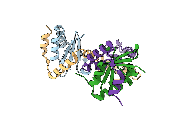 |
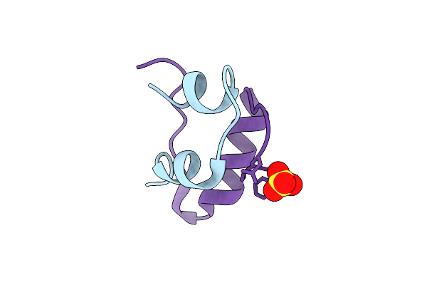
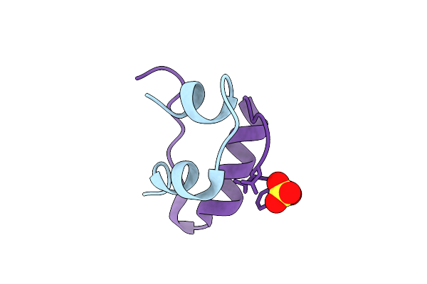
Mycobacterium Tuberculosis Relbe1 Toxin-Antitoxin System; Rv1247C (Relb1 Antitoxin), Rv1246C (Rele1 Toxin)
Organism: Mycobacterium tuberculosis h37rv
Method: X-RAY DIFFRACTION Release Date: 2025-12-17 Classification: TOXIN Ligands: CL |
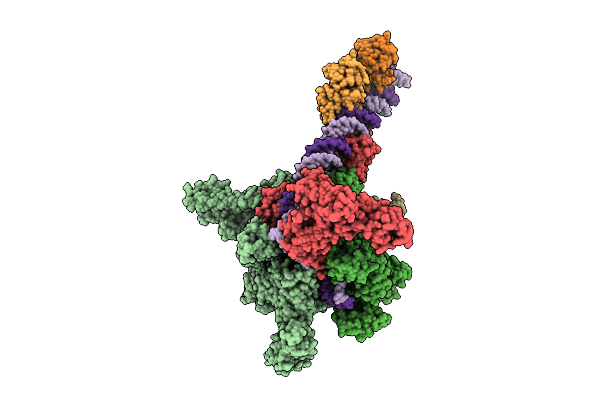 |
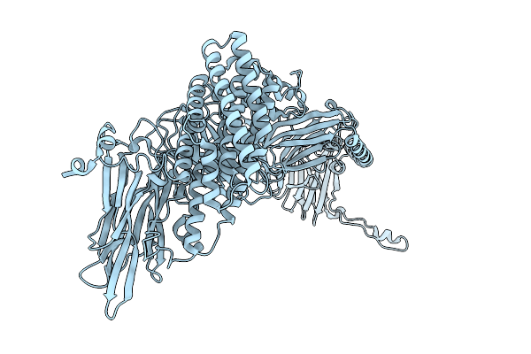
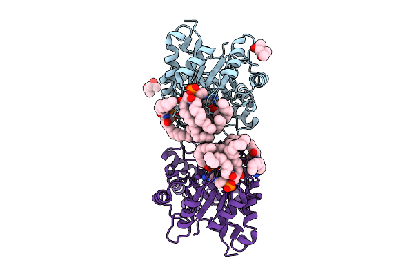
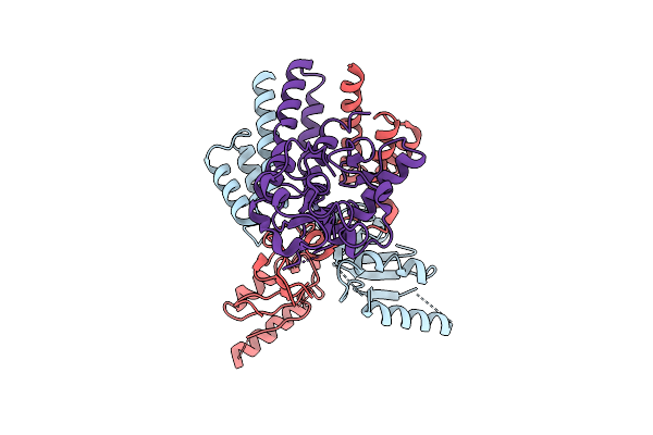
Cryo-Em Structure Of Vibrio Cholerae Rna Polymerase Transcription Activation Complex With Tcpp Transcription Factor And A Toxt Promoter Dna Fragment
Organism: Vibrio cholerae o395
Method: ELECTRON MICROSCOPY Release Date: 2025-12-17 Classification: TRANSCRIPTION Ligands: ZN, MG |
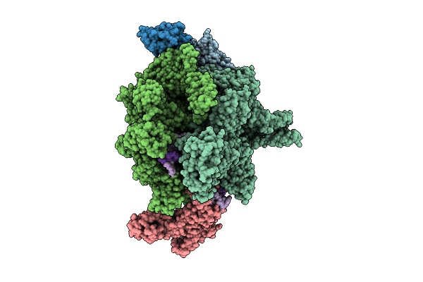 |
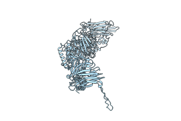
Cryo-Em Structure Of Vibrio Cholerae Rna Polymerase Transcription Activation Complex With Toxr And Tcpp Transcription Factors And A Toxt Promoter Dna Fragment
Organism: Vibrio cholerae o395, Vibrio cholerae o1 biovar el tor str. n16961
Method: ELECTRON MICROSCOPY Release Date: 2025-12-17 Classification: TRANSCRIPTION Ligands: MG, ZN |
 |
Organism: Streptomyces coelicolor
Method: ELECTRON MICROSCOPY Release Date: 2025-12-17 Classification: TOXIN Ligands: A2G, A1CAY, A1CAZ |
 |
Cryo-Em Structure Of Conivaptan-Bound Human Vasopressin V2 Receptor Complex With Fab
Organism: Homo sapiens, Escherichia coli
Method: ELECTRON MICROSCOPY Release Date: 2025-12-10 Classification: MEMBRANE PROTEIN/IMMUNE SYSTEM Ligands: A1ECE |
 |
Cryo-Em Structure Of Tolvaptan-Bound Human Vasopressin V2 Receptor Complex With Fab
Organism: Homo sapiens, Escherichia coli
Method: ELECTRON MICROSCOPY Release Date: 2025-12-10 Classification: MEMBRANE PROTEIN/IMMUNE SYSTEM Ligands: A1ECF |
 |
Native Structure Of The Full-Length Pesticidal Protein Cry8Ba2, From Crystals Formed In Vivo (Form 1)
Organism: Bacillus thuringiensis
Method: X-RAY DIFFRACTION Release Date: 2025-12-03 Classification: TOXIN |
 |
Native Structure Of The Full-Length Pesticidal Protein Cry8Ba2, From Crystals Formed In Vivo (Form 2)
Organism: Bacillus thuringiensis
Method: X-RAY DIFFRACTION Release Date: 2025-12-03 Classification: TOXIN |
 |
Organism: Homo sapiens
Method: X-RAY DIFFRACTION Release Date: 2025-12-03 Classification: TRANSCRIPTION Ligands: A1D7W |
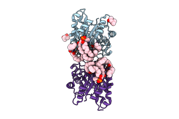 |
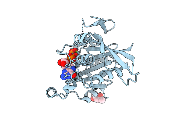
Structure Of Phospholipase D Betaib1I From Sicarius Terrosus Venom, H47N Mutant Bound To Product And Substrate Sphingolipids At 2.2 A Resolution From A 2-Day Old Crystal
Organism: Sicarius terrosus
Method: X-RAY DIFFRACTION Release Date: 2025-12-03 Classification: LYASE Ligands: A1A43, A1A44, NA, MG, MPD |
 |
Structure Of Phospholipase D Betaib1I From Sicarius Terrosus Venom, H47N Mutant Bound To Substrate Sphingolipids At 2.60 A Resolution
Organism: Sicarius terrosus
Method: X-RAY DIFFRACTION Release Date: 2025-12-03 Classification: LYASE Ligands: A1A43, NA, MG, MPD |
 |
Spitrobot-2 Advances Time-Resolvedcryo-Trapping Crystallography To Under 25 Ms: Human Insulin, Ph 9.0 (Apo State)
|
 |
Spitrobot-2 Advances Time-Resolvedcryo-Trapping Crystallography To Under 25 Ms: Human Insulin, Ph 4.5 (25 Ms Soaking)
Organism: Homo sapiens
Method: X-RAY DIFFRACTION Release Date: 2025-12-03 Classification: HORMONE Ligands: SO4 |
 |
Spitrobot-2 Advances Time-Resolvedcryo-Trapping Crystallography To Under 25 Ms: Human Insulin, Ph 4.5 (50 Ms Soaking)
Organism: Homo sapiens
Method: X-RAY DIFFRACTION Release Date: 2025-12-03 Classification: HORMONE Ligands: SO4 |
 |
Spitrobot-2 Advances Time-Resolvedcryo-Trapping Crystallography To Under 25 Ms: Human Insulin, Ph 4.5 (250 Ms Soaking)
Organism: Homo sapiens
Method: X-RAY DIFFRACTION Release Date: 2025-12-03 Classification: HORMONE Ligands: SO4 |
 |
Spitrobot-2 Advances Time-Resolvedcryo-Trapping Crystallography To Under 25 Ms: Human Insulin, Ph 4.5 (500 Ms Soaking)
Organism: Homo sapiens
Method: X-RAY DIFFRACTION Release Date: 2025-12-03 Classification: HORMONE Ligands: SO4 |
 |
Spitrobot-2 Advances Time-Resolvedcryo-Trapping Crystallography To Under 25 Ms: Human Insulin, Ph 4.5 (5 S Soaking)
Organism: Homo sapiens
Method: X-RAY DIFFRACTION Release Date: 2025-12-03 Classification: HORMONE Ligands: SO4 |
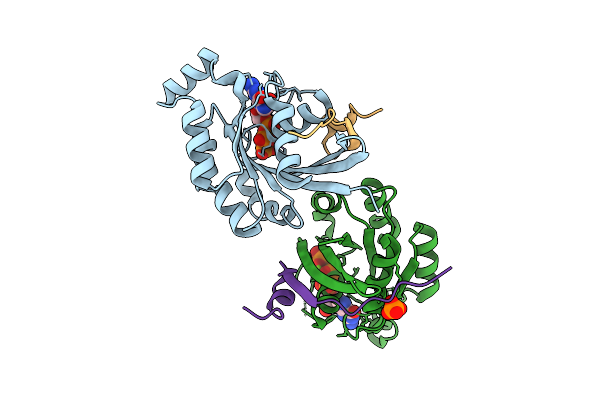 |
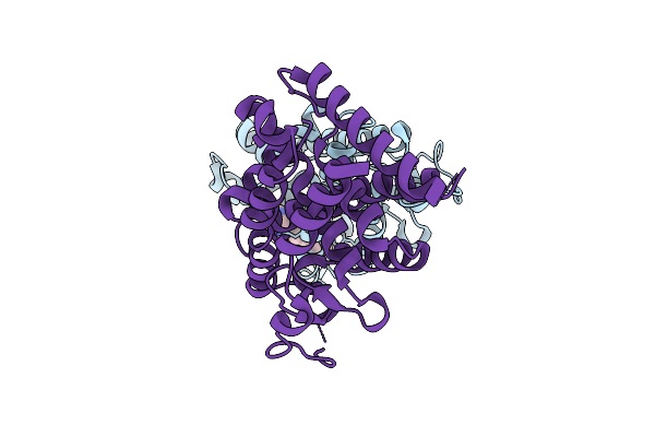
Crystal Structure Of Human Rac1 In Complex With The Scaffold Protein Posh (Residues 321-348)
Organism: Homo sapiens
Method: X-RAY DIFFRACTION Release Date: 2025-12-03 Classification: HYDROLASE Ligands: GNP, MG, PO4, CL |
 |
Crystal Structure Of Human Rac1 Fused With The Scaffold Protein Posh (Residues 319-371)
Organism: Homo sapiens
Method: X-RAY DIFFRACTION Release Date: 2025-12-03 Classification: HYDROLASE Ligands: GNP, MG, MPD |
 |
Organism: Escherichia coli
Method: ELECTRON MICROSCOPY Release Date: 2025-12-03 Classification: DNA BINDING PROTEIN Ligands: ZN |