
Search Count: 472
All
Selected
 |
Organism: Hepatitis c virus (isolate 1)
Method: SOLUTION NMR Release Date: 2026-01-28 Classification: RNA |
 |
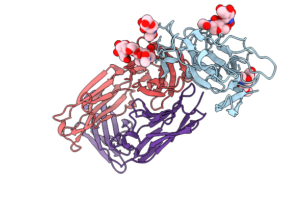
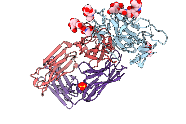
Crystal Structure Of Broadly Neutralizing Antibody Hepc108 In Complex With Hepatitis C Virus Envelope Glycoprotein E2 Ectodomain
Organism: Homo sapiens, Hepatitis c virus subtype 1b
Method: X-RAY DIFFRACTION Resolution:2.68 Å Release Date: 2026-01-21 Classification: IMMUNE SYSTEM Ligands: GOL, NAG |
 |
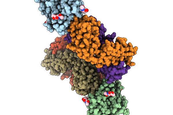
Structure Of Hepatitis C Virus Envelope Glycoprotein E2 Core From Genotype 6A Bound To Broadly Neutralizing Antibody Rm10-30
Organism: Macaca mulatta, Recombinant hepatitis c virus hk6a/jfh-1
Method: X-RAY DIFFRACTION Resolution:2.83 Å Release Date: 2026-01-14 Classification: VIRAL PROTEIN/IMMUNE SYSTEM Ligands: NAG |
 |
Structure Of Hepatitis C Virus Envelope Glycoprotein E2 Core From Genotype 6A Bound To Broadly Neutralizing Antibody Rm1-36
Organism: Macaca mulatta, Recombinant hepatitis c virus hk6a/jfh-1
Method: X-RAY DIFFRACTION Resolution:2.89 Å Release Date: 2026-01-14 Classification: VIRAL PROTEIN/IMMUNE SYSTEM Ligands: NAG |
 |
Structure Of Hepatitis C Virus Envelope Glycoprotein E2 Core From Genotype 6A Bound To Broadly Neutralizing Antibody Rm1-73
Organism: Macaca mulatta, Recombinant hepatitis c virus hk6a/jfh-1
Method: X-RAY DIFFRACTION Resolution:2.55 Å Release Date: 2026-01-14 Classification: VIRAL PROTEIN/IMMUNE SYSTEM Ligands: SO4, NAG |
 |
Structure Of Hepatitis C Virus Envelope Glycoprotein E2 Core From Genotype 6A Bound To Broadly Neutralizing Antibody Rm11-48
Organism: Recombinant hepatitis c virus hk6a/jfh-1, Macaca mulatta
Method: X-RAY DIFFRACTION Resolution:2.85 Å Release Date: 2026-01-14 Classification: VIRAL PROTEIN/IMMUNE SYSTEM Ligands: NAG, SO4 |
 |
Structure Of Hepatitis C Virus Envelope Glycoprotein Hcv-1 E2Ecto From Genotype 1A Bound To Neutralizing Antibody Rm5-16
Organism: Macaca mulatta, Homo sapiens, Hepatitis c virus (isolate 1)
Method: X-RAY DIFFRACTION Resolution:3.39 Å Release Date: 2026-01-14 Classification: VIRAL PROTEIN/IMMUNE SYSTEM Ligands: NAG, MAN |
 |
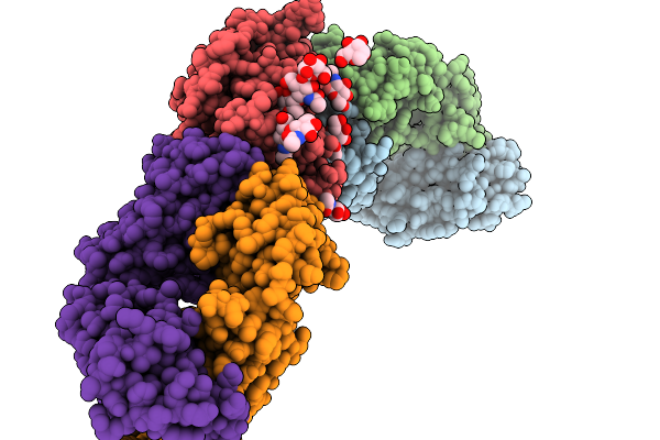
Structural Insights Into The Polymerase Catalyzed Fad-Capping Of Hepatitis C Viral Rna
Organism: Hepatitis c virus jfh-1, Synthetic construct
Method: X-RAY DIFFRACTION Resolution:2.74 Å Release Date: 2025-08-13 Classification: VIRAL PROTEIN Ligands: CDP, FAD, MN |
 |
Structural Insights Into The Polymerase Catalyzed Fad-Capping Of Hepatitis C Viral Rna
Organism: Hepatitis c virus jfh-1, Synthetic construct
Method: X-RAY DIFFRACTION Resolution:2.96 Å Release Date: 2025-08-13 Classification: VIRAL PROTEIN Ligands: FAD, CDP, MN |
 |
Structural Insights Into The Polymerase Catalyzed Fad-Capping Of Hepatitis C Viral Rna
Organism: Hepatitis c virus jfh-1, Synthetic construct
Method: X-RAY DIFFRACTION Resolution:3.46 Å Release Date: 2025-08-13 Classification: VIRAL PROTEIN Ligands: CDP, MN, FAD |
 |
Structural Insights Into The Polymerase Catalyzed Fad-Capping Of Hepatitis C Viral Rna
Organism: Hepatitis c virus jfh-1, Synthetic construct
Method: X-RAY DIFFRACTION Resolution:2.92 Å Release Date: 2025-08-13 Classification: VIRAL PROTEIN Ligands: FAD, GDP, MN |
 |
Structural Insights Into The Polymerase Catalyzed Fad-Capping Of Hepatitis C Viral Rna
Organism: Hepatitis c virus jfh-1, Synthetic construct
Method: X-RAY DIFFRACTION Resolution:2.25 Å Release Date: 2025-08-13 Classification: VIRAL PROTEIN Ligands: C5P, MN, FAD |
 |
Structural Insights Into The Polymerase Catalyzed Fad-Capping Of Hepatitis C Viral Rna
Organism: Hepatitis c virus genotype 2a (isolate jfh-1), Synthetic construct
Method: X-RAY DIFFRACTION Resolution:3.13 Å Release Date: 2025-08-13 Classification: VIRAL PROTEIN Ligands: GDP, MN, FAD |
 |
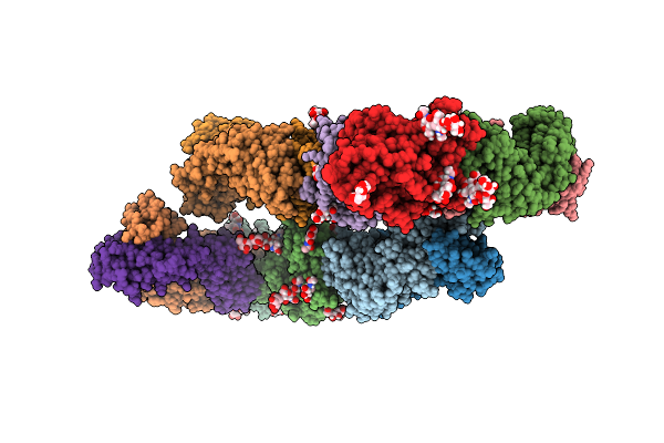
Cryoem Structure Of Non-Neutralizing Bivalent Antibody Cbh-4B In Complex With Hepatitis C Virus Envelope Glycoprotein E2
Organism: Homo sapiens, Hepatitis c virus (isolate 1)
Method: ELECTRON MICROSCOPY Release Date: 2024-09-04 Classification: VIRAL PROTEIN/IMMUNE SYSTEM |
 |
Cryoem Structure Of Non-Neutralizing Antibody Cbh-4B In Complex With Hepatitis C Virus Envelope Glycoprotein E2
Organism: Homo sapiens, Hepatitis c virus (isolate 1)
Method: ELECTRON MICROSCOPY Release Date: 2024-08-28 Classification: ANTIVIRAL PROTEIN |
 |
Structure Of Hepatitis C Virus Envelope N-Terminal Truncated Glycoprotein 2 (E2) (Residues 456-713) From J6 Genotype
Organism: Hepatitis c virus isolate hc-j6, Mus musculus
Method: X-RAY DIFFRACTION Resolution:2.45 Å Release Date: 2023-03-29 Classification: VIRAL PROTEIN |
 |
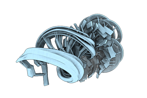
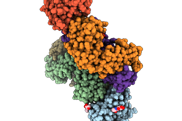
Structure Of The Hcv Ires Bound To The 40S Ribosomal Subunit, Head Open. Structure 9(Delta Dii)
Organism: Hepatitis c virus (isolate 1), Oryctolagus cuniculus
Method: ELECTRON MICROSCOPY Release Date: 2022-07-27 Classification: RIBOSOME Ligands: ZN |
 |
Structure Of The Wt Ires And 40S Ribosome Binary Complex, Open Conformation. Structure 10(Wt)
Organism: Hepatitis c virus (isolate 1), Oryctolagus cuniculus
Method: ELECTRON MICROSCOPY Release Date: 2022-07-27 Classification: RIBOSOME Ligands: ZN |
 |
Structure Of The Wt Ires And 40S Ribosome Ternary Complex, Open Conformation. Structure 11(Wt)
Organism: Homo sapiens, Hepatitis c virus (isolate 1), Oryctolagus cuniculus
Method: ELECTRON MICROSCOPY Release Date: 2022-07-27 Classification: RIBOSOME Ligands: ZN |
 |
Structure Of The Wt Ires Eif2-Containing 48S Initiation Complex, Closed Conformation. Structure 12(Wt).
Organism: Homo sapiens, Hepatitis c virus (isolate 1), Oryctolagus cuniculus
Method: ELECTRON MICROSCOPY Release Date: 2022-07-27 Classification: RIBOSOME Ligands: ZN |