
Search Count: 16
 |
Organism: Mangifera indica
Method: X-RAY DIFFRACTION Resolution:2.40 Å Release Date: 2024-08-07 Classification: PLANT PROTEIN Ligands: PG4 |
 |
Organism: Klebsiella pneumoniae mgh 78578
Method: X-RAY DIFFRACTION Resolution:2.80 Å Release Date: 2020-02-26 Classification: TRANSFERASE Ligands: EDO, CYS, PO4 |
 |
The Ultra High Resolution Structure Of A Novel Alpha-L-Arabinofuranosidase (Ctgh43) From Clostridium Thermocellum Atcc 27405 With Bound Trimethyl N-Oxide (Trs)
Organism: Clostridium thermocellum
Method: X-RAY DIFFRACTION Resolution:0.97 Å Release Date: 2016-07-27 Classification: HYDROLASE Ligands: CA, TRS |
 |
The High Resolution Structure Of A Novel Alpha-L-Arabinofuranosidase (Ctgh43) From Clostridium Thermocellum Atcc 27405
Organism: Clostridium thermocellum
Method: X-RAY DIFFRACTION Resolution:1.65 Å Release Date: 2016-07-27 Classification: HYDROLASE Ligands: SO4, ACT, GOL |
 |
Organism: Bacillus anthracis
Method: X-RAY DIFFRACTION Resolution:1.87 Å Release Date: 2013-11-13 Classification: TOXIN Ligands: SO4 |
 |
Organism: Homo sapiens
Method: X-RAY DIFFRACTION Resolution:2.01 Å Release Date: 2013-10-09 Classification: LYASE Ligands: FES, CHD, SAL, EDO |
 |
Organism: Mycobacterium tuberculosis
Method: X-RAY DIFFRACTION Resolution:3.00 Å Release Date: 2011-04-27 Classification: METAL BINDING PROTEIN |
 |
Structural Ordering Of Disordered Ligand Binding Loops Of Biotin Protein Ligase Into Active Conformations As A Consequence Of Dehydration
Organism: Mycobacterium tuberculosis
Method: X-RAY DIFFRACTION Resolution:2.69 Å Release Date: 2010-03-09 Classification: LIGASE |
 |
Organism: Mycobacterium tuberculosis
Method: X-RAY DIFFRACTION Resolution:2.80 Å Release Date: 2010-03-09 Classification: LIGASE |
 |
Organism: Mycobacterium tuberculosis
Method: X-RAY DIFFRACTION Resolution:2.59 Å Release Date: 2009-12-15 Classification: METAL BINDING PROTEIN Ligands: FE, UNX, UNL |
 |
Organism: Homo sapiens
Method: X-RAY DIFFRACTION Resolution:1.90 Å Release Date: 2004-08-17 Classification: IMMUNE SYSTEM Ligands: CD |
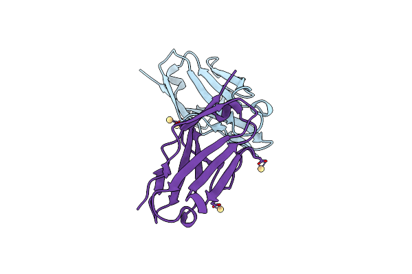 |
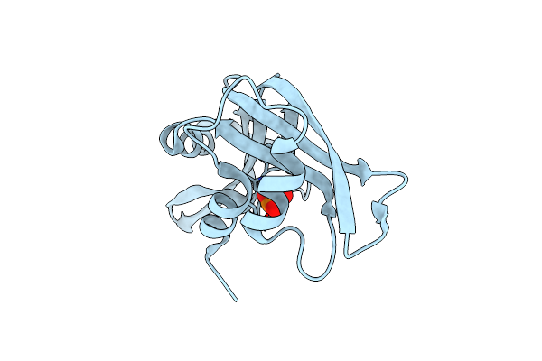
High Resolution Crystal Structure Of Jto2, A Mutant Of The Non-Amyloidogenic Lamba6 Light Chain, Jto
Organism: Homo sapiens
Method: X-RAY DIFFRACTION Resolution:1.60 Å Release Date: 2004-07-13 Classification: IMMUNE SYSTEM Ligands: CD |
 |
Immunoglobulin Motif Dna-Recognition And Heterodimerization For The Pebp2/Cbf Runt-Domain
|
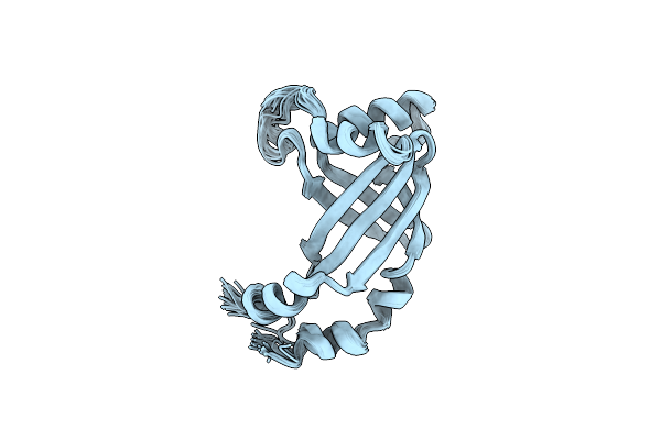 |
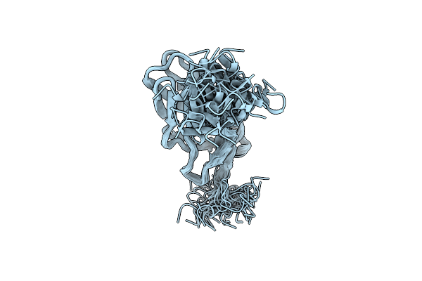
Molecular Insights Into Pebp2/Cbf-Smmhc Associated Acute Leukemia Revealed From The Three-Dimensional Structure Of Pebp2/Cbf Beta
Organism: Homo sapiens
Method: SOLUTION NMR Release Date: 2000-01-01 Classification: GENE REGULATION |
 |
Organism: Rattus norvegicus
Method: X-RAY DIFFRACTION Resolution:2.50 Å Release Date: 1998-12-09 Classification: HYDROLASE Ligands: PO4 |
 |
Structure Of The Crystalline Complex Of Cytidylic Acid (2'-Cmp) With Ribonuclease At 1.6 Angstroms Resolution
Organism: Bos taurus
Method: X-RAY DIFFRACTION Resolution:1.60 Å Release Date: 1994-01-31 Classification: HYDROLASE(ENDORIBONUCLEASE) Ligands: C2P |