
Search Count: 46
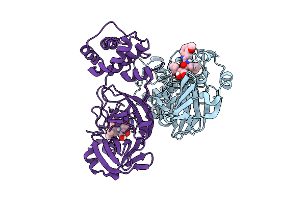 |
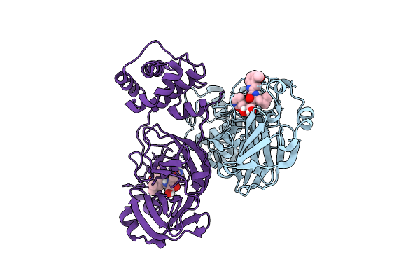
Crystal Structure Of Sars-Cov-2 Mpro S10A In Complex With Pfizer Intravenous Inhibitor Pf-00835231
Organism: Severe acute respiratory syndrome coronavirus 2
Method: X-RAY DIFFRACTION Release Date: 2025-12-10 Classification: VIRAL PROTEIN Ligands: V2M |
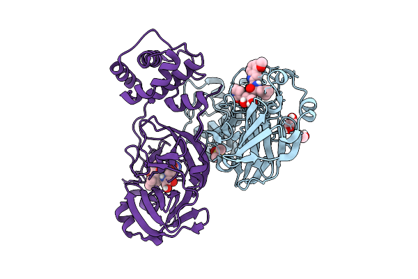 |
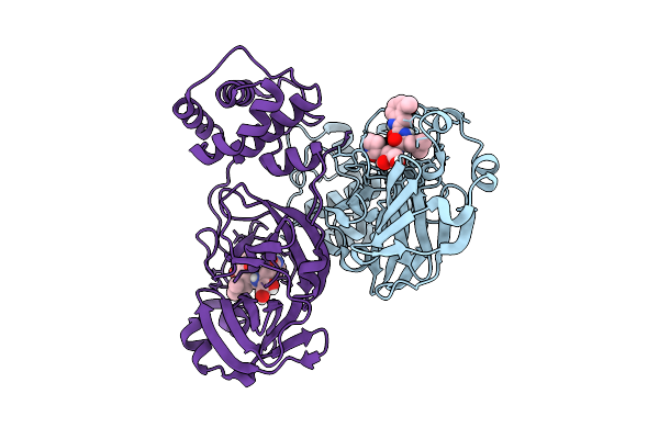
Crystal Structure Of Sars-Cov-2 Mpro S10C In Complex With Pfizer Intravenous Inhibitor Pf-00835231
Organism: Severe acute respiratory syndrome coronavirus 2
Method: X-RAY DIFFRACTION Release Date: 2025-12-10 Classification: VIRAL PROTEIN Ligands: DMS, EDO, V2M |
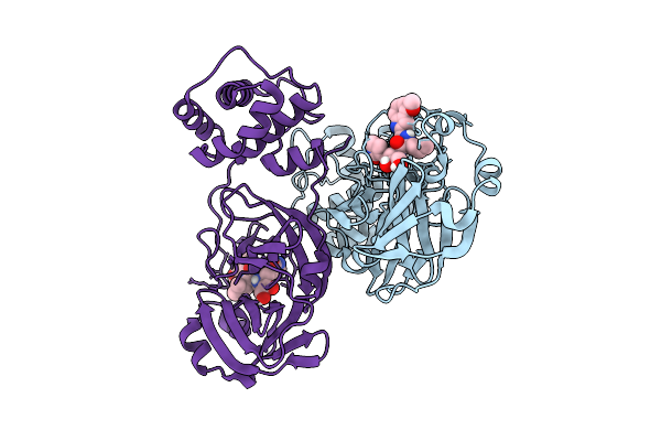 |
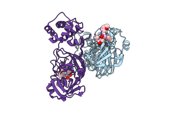
Crystal Structure Of Sars-Cov-2 Mpro S113A In Complex With Pfizer Intravenous Inhibitor Pf-00835231
Organism: Severe acute respiratory syndrome coronavirus 2
Method: X-RAY DIFFRACTION Release Date: 2025-12-10 Classification: VIRAL PROTEIN Ligands: V2M |
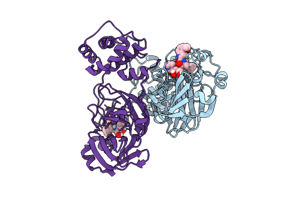 |
Crystal Structure Of Sars-Cov-2 Mpro S113C In Complex With Pfizer Intravenous Inhibitor Pf-00835231
Organism: Severe acute respiratory syndrome coronavirus 2
Method: X-RAY DIFFRACTION Release Date: 2025-12-10 Classification: VIRAL PROTEIN Ligands: V2M |
 |
Crystal Structure Of Sars-Cov-2 Mpro L115A In Complex With Pfizer Intravenous Inhibitor Pf-00835231
Organism: Severe acute respiratory syndrome coronavirus 2
Method: X-RAY DIFFRACTION Release Date: 2025-12-10 Classification: VIRAL PROTEIN Ligands: V2M |
 |
Crystal Structure Of Sars-Cov-2 Mpro L115M In Complex With Pfizer Intravenous Inhibitor Pf-00835231
Organism: Severe acute respiratory syndrome coronavirus 2
Method: X-RAY DIFFRACTION Release Date: 2025-12-10 Classification: VIRAL PROTEIN Ligands: V2M |
 |
Crystal Structure Of Sars-Cov-2 Mpro S147A In Complex With Pfizer Intravenous Inhibitor Pf-00835231
Organism: Severe acute respiratory syndrome coronavirus 2
Method: X-RAY DIFFRACTION Release Date: 2025-12-10 Classification: VIRAL PROTEIN Ligands: V2M |
 |
Crystal Structure Of Sars-Cov-2 Mpro S147N In Complex With Pfizer Intravenous Inhibitor Pf-00835231
Organism: Severe acute respiratory syndrome coronavirus 2
Method: X-RAY DIFFRACTION Release Date: 2025-12-10 Classification: VIRAL PROTEIN Ligands: V2M |
 |
Organism: Henipavirus nipahense
Method: ELECTRON MICROSCOPY Release Date: 2025-07-30 Classification: TRANSFERASE |
 |
Cryo-Em Structure Of The Nipah Virus Polymerase Containing The Connecting Domain
Organism: Henipavirus nipahense
Method: ELECTRON MICROSCOPY Release Date: 2025-07-30 Classification: VIRAL PROTEIN, TRANSFERASE |
 |
Organism: Henipavirus nipahense
Method: ELECTRON MICROSCOPY Release Date: 2025-07-23 Classification: VIRAL PROTEIN, TRANSFERASE |
 |
Organism: Homo sapiens
Method: ELECTRON MICROSCOPY Release Date: 2024-05-01 Classification: STRUCTURAL PROTEIN Ligands: GTP, MG, GDP |
 |
Crystal Structure Of Haloacid Dehalogenase-Like Hydrolase Family Enzyme From Staphylococcus Lugdunensis
Organism: Staphylococcus lugdunensis
Method: X-RAY DIFFRACTION Resolution:1.73 Å Release Date: 2023-12-27 Classification: HYDROLASE Ligands: PEG, EDO, OXM, FMT |
 |
Crystal Structure Of Putative Amino Acid Binding Periplasmic Abc Transporter Protein From Candidatus Liberibacter Asiaticus In Complex With Clidinium
Organism: Liberibacter asiaticus (strain psy62)
Method: X-RAY DIFFRACTION Resolution:2.56 Å Release Date: 2023-07-26 Classification: TRANSPORT PROTEIN Ligands: KG2 |
 |
Crystal Structure Of Putative Amino Acid Binding Periplasmic Abc Transporter Protein From Candidatus Liberibacter Asiaticus In Complex With Pimozide
Organism: Candidatus liberibacter asiaticus str. psy62
Method: X-RAY DIFFRACTION Resolution:2.65 Å Release Date: 2023-07-26 Classification: TRANSPORT PROTEIN Ligands: 1II |
 |
Organism: Homo sapiens
Method: X-RAY DIFFRACTION Resolution:1.45 Å Release Date: 2023-06-21 Classification: TRANSFERASE/INHIBITOR Ligands: I6C, GOL |
 |
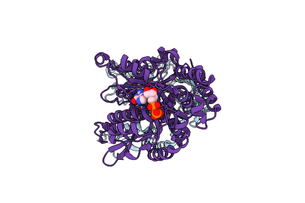
Organism: Homo sapiens
Method: X-RAY DIFFRACTION Resolution:2.50 Å Release Date: 2021-10-13 Classification: OXYGEN TRANSPORT Ligands: CMO, HEM |
 |
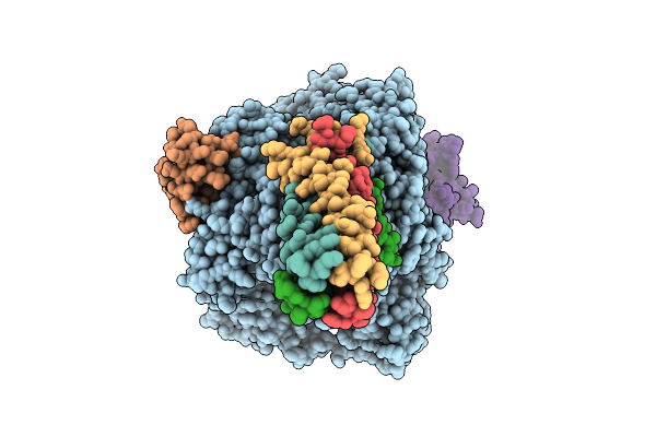
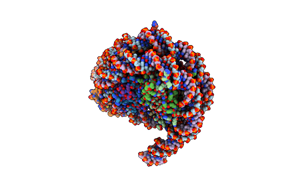
Organism: Xenopus laevis, Xenopus tropicalis, Homo sapiens
Method: ELECTRON MICROSCOPY Release Date: 2020-10-21 Classification: GENE REGULATION Ligands: SAM, ZN |
 |
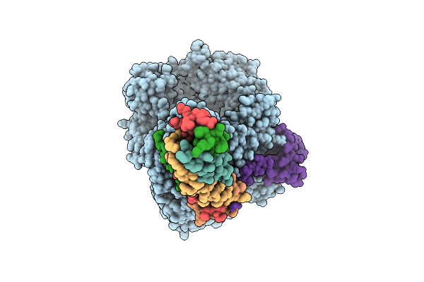
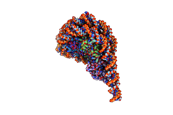
Nsd3 Bearing E1181K/T1232A Dual Mutation In Complex With 187-Bp Ncp (1:1 Binding Mode)
Organism: Xenopus laevis, Xenopus tropicalis, Homo sapiens
Method: ELECTRON MICROSCOPY Release Date: 2020-10-21 Classification: GENE REGULATION Ligands: SAM, ZN |
 |
Nsd3 Bearing E1181K/T1232A Dual Mutation In Complex With 187-Bp Ncp (2:1 Binding Mode)
Organism: Xenopus laevis, Xenopus tropicalis, Homo sapiens
Method: ELECTRON MICROSCOPY Release Date: 2020-10-21 Classification: GENE REGULATION Ligands: SAM, ZN |