
Search Count: 683
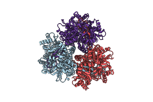 |
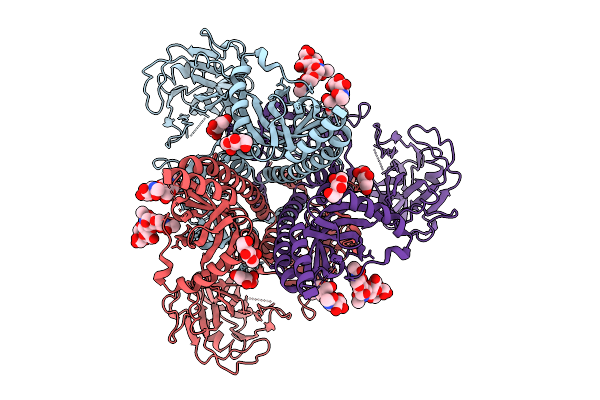
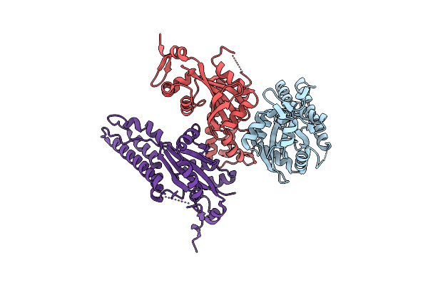
Structure Of Acrb In The Form Of Native Cell Membrane Nanoparticles (Ncmnp33-50)
Organism: Escherichia coli
Method: ELECTRON MICROSCOPY Release Date: 2025-10-15 Classification: MEMBRANE PROTEIN |
 |
Crystal Structure Of Aedes Aegypti Dopachrome Conversion Enzyme With L-Dopamine.
Organism: Aedes aegypti
Method: X-RAY DIFFRACTION Release Date: 2025-09-24 Classification: ISOMERASE Ligands: CA, ACY, LDP, NDG |
 |
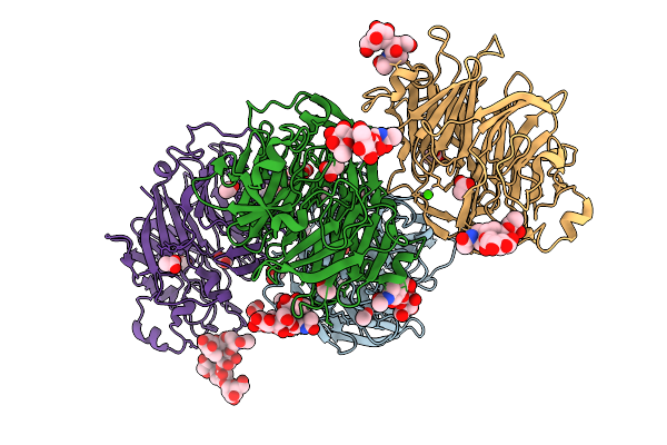
Cryo-Em Structure Of Neutralizing Murine Antibody Ws.Hsv-1.24 In Complex With Hsv-1 Glycoprotein B Trimer Gb-Ecto.516P.531E
Organism: Human alphaherpesvirus 1, Mus musculus
Method: ELECTRON MICROSCOPY Release Date: 2025-09-10 Classification: VIRAL PROTEIN Ligands: NAG |
 |
Cryo-Em Structure Of Neutralizing Human Antibody D48 In Complex With Hsv-1 Glycoprotein B Trimer Gb-Ecto.516P.531E.Ds
Organism: Human alphaherpesvirus 1, Homo sapiens
Method: ELECTRON MICROSCOPY Release Date: 2025-09-10 Classification: VIRAL PROTEIN Ligands: NAG |
 |
Cryo-Em Structure Of Neutralizing Human Antibody D48 In Complex With Hsv-1 Glycoprotein B Trimer Gb-Ecto.516P
Organism: Human alphaherpesvirus 1, Homo sapiens
Method: ELECTRON MICROSCOPY Release Date: 2025-09-10 Classification: VIRAL PROTEIN Ligands: NAG |
 |
Cryo-Em Structure Of Gb-Ecto.516P.531E.Ds, A Prefusion-Stabilized Hsv-1 Glycoprotein B Extracellular Domain
Organism: Human alphaherpesvirus 1
Method: ELECTRON MICROSCOPY Release Date: 2025-09-10 Classification: VIRAL PROTEIN Ligands: NAG |
 |
Cryo-Em Structure Of Gb-Ecto.516P, An Hsv-1 Glycoprotein B Extracellular Domain
Organism: Human alphaherpesvirus 1
Method: ELECTRON MICROSCOPY Release Date: 2025-09-10 Classification: VIRAL PROTEIN Ligands: NAG |
 |
Crystal Structure Of Sars-Cov-2 Rbd In Complex With A Neutralizing Antibody Scfv N1
Organism: Homo sapiens, Severe acute respiratory syndrome coronavirus 2
Method: X-RAY DIFFRACTION Release Date: 2025-09-03 Classification: VIRAL PROTEIN/IMMUNE SYSTEM Ligands: NAG |
 |
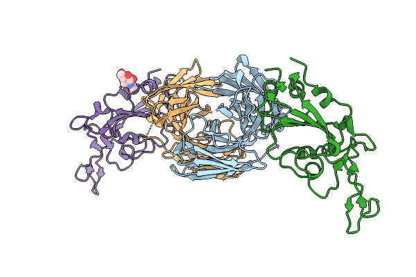
The Glycoprotein E Of Varicella-Zoster Virus In Complex With Wll-1/Wll-28 Fab
Organism: Varicella-zoster virus (strain oka vaccine), Homo sapiens
Method: ELECTRON MICROSCOPY Release Date: 2025-09-03 Classification: VIRAL PROTEIN/IMMUNE SYSTEM |
 |
Organism: Aedes aegypti
Method: X-RAY DIFFRACTION Release Date: 2025-09-03 Classification: ISOMERASE Ligands: CA, ACY |
 |
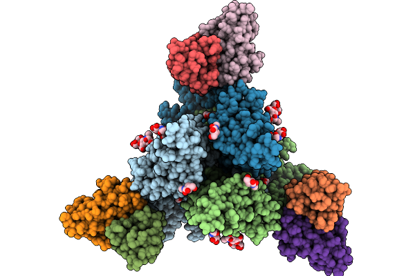
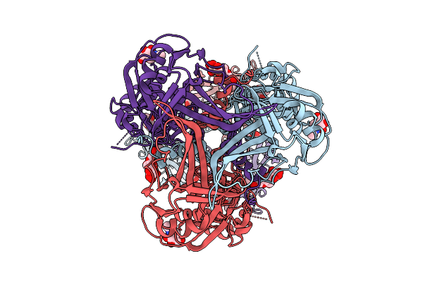
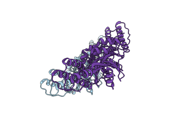
Crystal Structure Of Escherichia Coli Groel With Magnesium Ions And A Phosphorylated Serine Residue-3.2A
Organism: Escherichia coli bl21(de3)
Method: X-RAY DIFFRACTION Release Date: 2025-09-03 Classification: CHAPERONE Ligands: HEZ, MG, BME, TAM |
 |
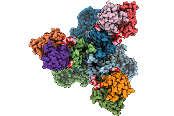
Crystal Structure Of Escherichia Coli Groel With Magnesium Ions And A Phosphorylated Serine Residue-3.0 A
Organism: Escherichia coli bl21(de3)
Method: X-RAY DIFFRACTION Release Date: 2025-09-03 Classification: CHAPERONE Ligands: BME, HEZ, MG |
 |
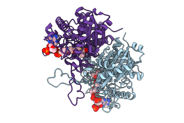
Organism: Burkholderia sp. rf2-non_bp3
Method: X-RAY DIFFRACTION Release Date: 2025-08-27 Classification: LYASE |
 |
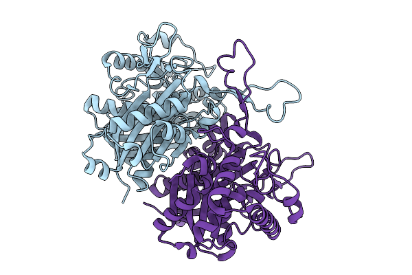
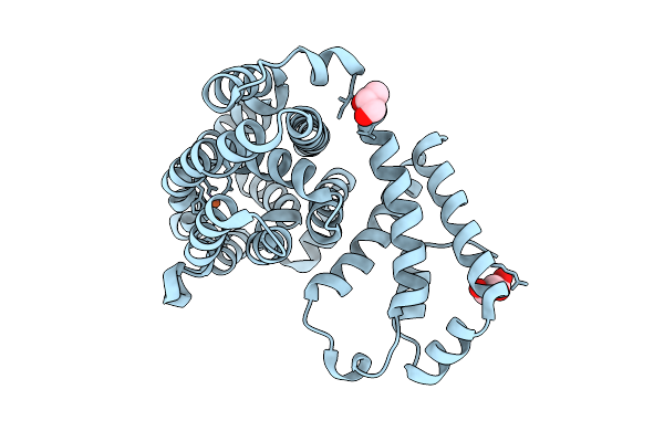
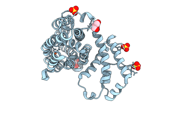
Crystal Structure Of Polyketoacyl-Coa Thiolase From Burkholderia Sp In Complex With Butyryl-Coa
Organism: Burkholderia sp. rf2-non_bp3
Method: X-RAY DIFFRACTION Release Date: 2025-08-27 Classification: LYASE Ligands: COA |
 |
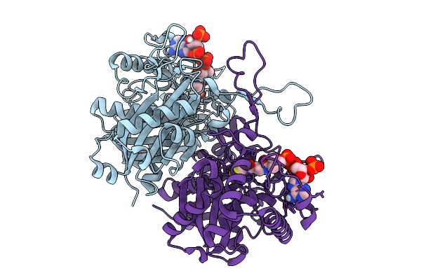
Crystal Structure Of Polyketoacyl-Coa Thiolase From Burkholderia Sp. In Complex With Acetyl-Coa
Organism: Burkholderia sp. rf2-non_bp3
Method: X-RAY DIFFRACTION Release Date: 2025-08-27 Classification: LYASE Ligands: COA |
 |
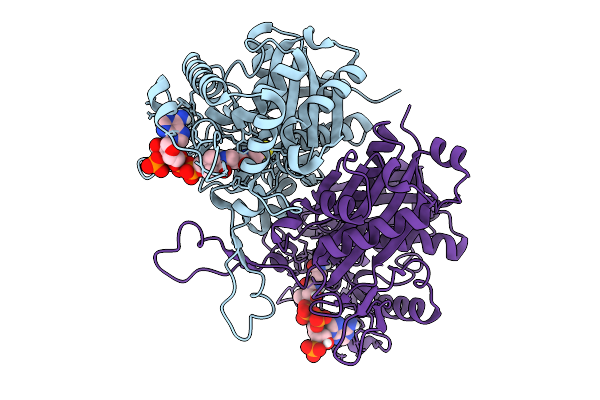
Crystal Structure Of Polyketoacyl-Coa Thiolase From Burkholderia Sp. In Complex With Acetoacetyl-Coa
Organism: Burkholderia sp. rf2-non_bp3
Method: X-RAY DIFFRACTION Release Date: 2025-08-27 Classification: LYASE Ligands: COA, A1BNQ |
 |
Organism: Human respiratory syncytial virus a
Method: X-RAY DIFFRACTION Release Date: 2025-08-20 Classification: VIRAL PROTEIN |
 |
Organism: Respiratory syncytial virus
Method: X-RAY DIFFRACTION Release Date: 2025-08-20 Classification: VIRAL PROTEIN |
 |
Organism: Streptomyces griseoflavus
Method: X-RAY DIFFRACTION Release Date: 2025-08-20 Classification: OXIDOREDUCTASE Ligands: FE, GOL |
 |
Crystal Structure Of N-Oxygenase Hrmi With The Diferric Cofactor Partially Loaded
Organism: Streptomyces griseoflavus
Method: X-RAY DIFFRACTION Release Date: 2025-08-20 Classification: OXIDOREDUCTASE Ligands: FE, SO4, GOL |