
Search Count: 26
 |
Organism: Mangifera indica
Method: X-RAY DIFFRACTION Release Date: 2024-12-18 Classification: TRANSFERASE Ligands: UDP |
 |
Organism: Mangifera indica
Method: X-RAY DIFFRACTION Release Date: 2024-12-18 Classification: TRANSFERASE Ligands: UDP |
 |
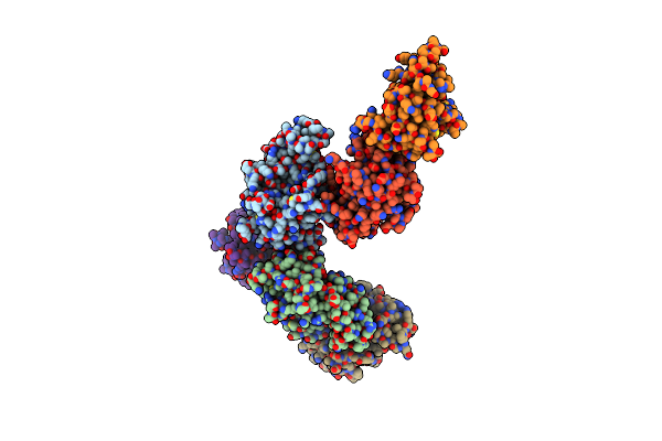
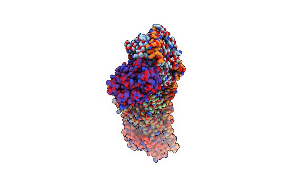
Organism: Homo sapiens
Method: ELECTRON MICROSCOPY Release Date: 2024-08-14 Classification: CHAPERONE |
 |
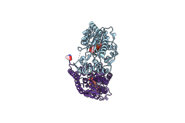
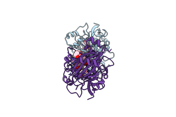
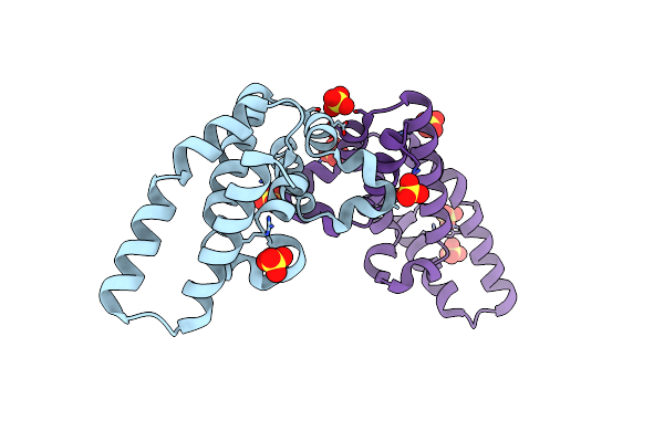
Oomycete Nudix Effectors Display Wy-Nudix Conformations With Mrna Decapping Activity
Organism: Phytophthora sojae strain p6497
Method: X-RAY DIFFRACTION Resolution:3.19 Å Release Date: 2024-07-03 Classification: HYDROLASE |
 |
Organism: Bacillus subtilis
Method: X-RAY DIFFRACTION Resolution:1.90 Å Release Date: 2022-10-12 Classification: TRANSFERASE Ligands: UDP, GOL, TRS |
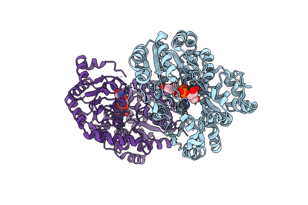 |
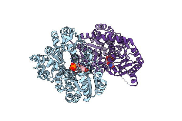
Organism: Mangifera indica
Method: X-RAY DIFFRACTION Resolution:2.85 Å Release Date: 2022-07-06 Classification: TRANSFERASE Ligands: UPG |
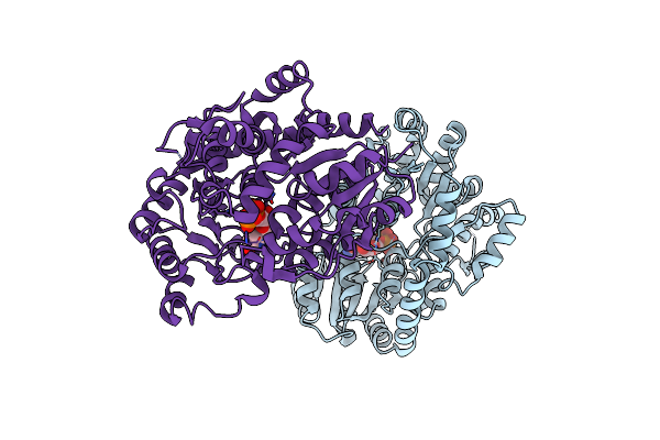 |
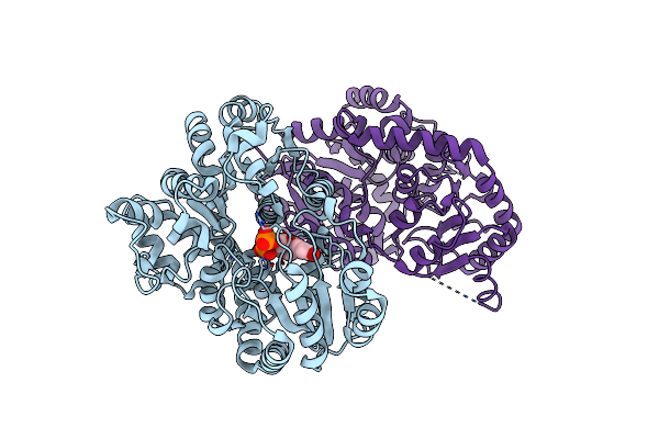
Organism: Mangifera indica
Method: X-RAY DIFFRACTION Resolution:3.10 Å Release Date: 2022-07-06 Classification: TRANSFERASE Ligands: UDP |
 |
Crystal Structures Of 2-Oxoglutarate Dependent Dioxygenase (Ctb9) From Cercospora Sp. Jnu001
Organism: Cercospora sojina
Method: X-RAY DIFFRACTION Resolution:2.30 Å Release Date: 2022-05-25 Classification: OXIDOREDUCTASE Ligands: CU, GOL |
 |
Crystal Structures Of 2-Oxoglutarate Dependent Dioxygenase (Ctb9) In Complex With N-Oxalylglycine
Organism: Cercospora sojina
Method: X-RAY DIFFRACTION Resolution:2.50 Å Release Date: 2022-05-25 Classification: OXIDOREDUCTASE Ligands: CU, OGA, EDO, GOL |
 |
Crystal Structures Of 2-Oxoglutarate Dependent Dioxygenase (Ctb9) In Complex With N-Oxalylglycine And Pre-Cercosporin
Organism: Cercospora sojina
Method: X-RAY DIFFRACTION Resolution:2.20 Å Release Date: 2022-05-25 Classification: OXIDOREDUCTASE Ligands: CU, OGA, JD9, EDO |
 |
Discovery Of Novel Small-Molecule Inhibitors Of Pd-1/Pd-L1 Axis That Promotes Pd-L1 Internalization And Degradation
Organism: Homo sapiens
Method: X-RAY DIFFRACTION Resolution:2.42 Å Release Date: 2022-01-26 Classification: IMMUNOSUPPRESSANT Ligands: HOU |
 |
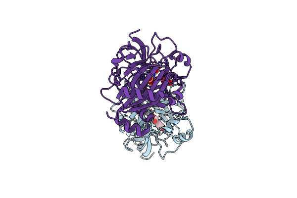
Organism: Phytophthora sojae (strain p6497), Glycine soja
Method: X-RAY DIFFRACTION Resolution:2.51 Å Release Date: 2021-03-17 Classification: IMMUNE SYSTEM |
 |
Organism: Rattus norvegicus, Gallus gallus, Bos taurus
Method: X-RAY DIFFRACTION Resolution:2.00 Å Release Date: 2020-07-08 Classification: CELL CYCLE Ligands: GTP, MG, GOL, GDP, L95, MES, ACP |
 |
Substrates Promiscuity Of Xyloglucanases And Endoglucanases Of Glycoside Hydrolase 12 Family
Organism: Aspergillus fischeri
Method: X-RAY DIFFRACTION Resolution:2.03 Å Release Date: 2020-06-17 Classification: HYDROLASE |
 |
Organism: Aspergillus fischeri
Method: X-RAY DIFFRACTION Resolution:1.51 Å Release Date: 2020-06-17 Classification: HYDROLASE |
 |
Crystal Structure Of Bacterial Pirin Yhhw In Complex With Nickel(Ii) From Escherichia Coli
Organism: Escherichia coli (strain k12)
Method: X-RAY DIFFRACTION Resolution:3.09 Å Release Date: 2019-11-06 Classification: METAL BINDING PROTEIN Ligands: NI |
 |
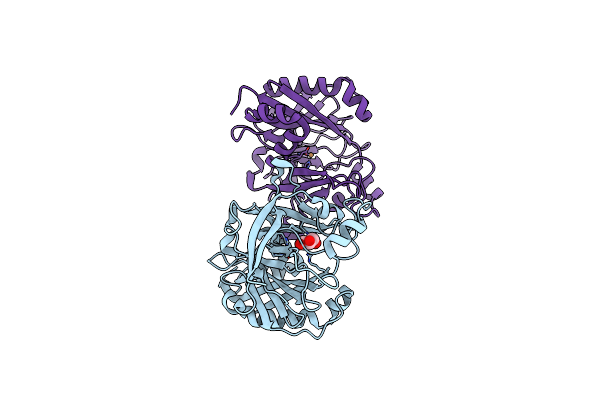
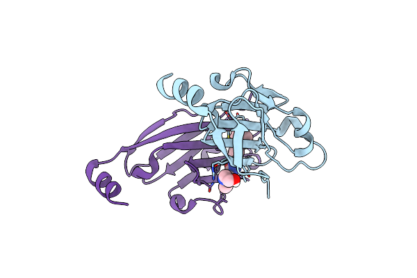
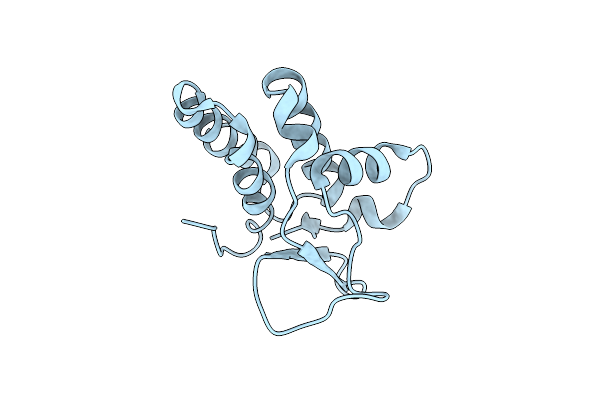
Phytophthora Sojae Effector Psavh240 Inhibits A Host Aspartic Protease Secretion To Promote Infection
Organism: Phytophthora sojae
Method: X-RAY DIFFRACTION Resolution:2.30 Å Release Date: 2019-02-06 Classification: IMMUNE SYSTEM Ligands: SO4 |
 |
Organism: Phytophthora sojae
Method: X-RAY DIFFRACTION Resolution:2.80 Å Release Date: 2017-08-16 Classification: UNKNOWN FUNCTION |
 |
 |
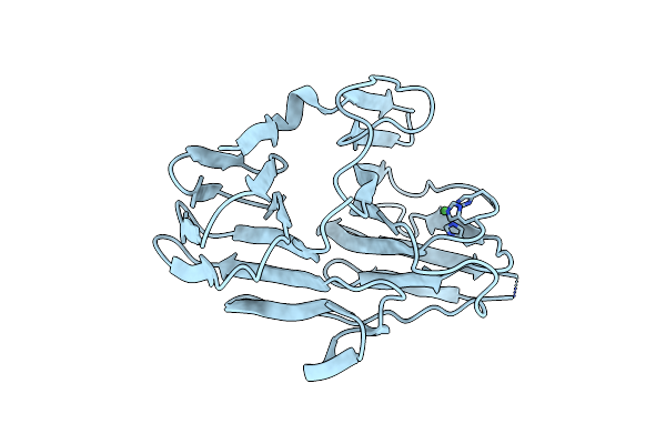
Structure Of The Autocatalytic Cysteine Protease Domain Of Potyvirus Helper-Component Proteinase
Organism: Turnip mosaic virus
Method: X-RAY DIFFRACTION Resolution:2.00 Å Release Date: 2011-05-04 Classification: HYDROLASE |