Search Count: 57
 |
Organism: Homo sapiens
Method: X-RAY DIFFRACTION Resolution:2.34 Å Release Date: 2024-01-24 Classification: OXIDOREDUCTASE Ligands: NAI, YBO, EDO, PEG |
 |
Organism: Homo sapiens
Method: X-RAY DIFFRACTION Resolution:2.97 Å Release Date: 2021-07-14 Classification: OXIDOREDUCTASE Ligands: EDO, PO4, PEG, GOL, ATP |
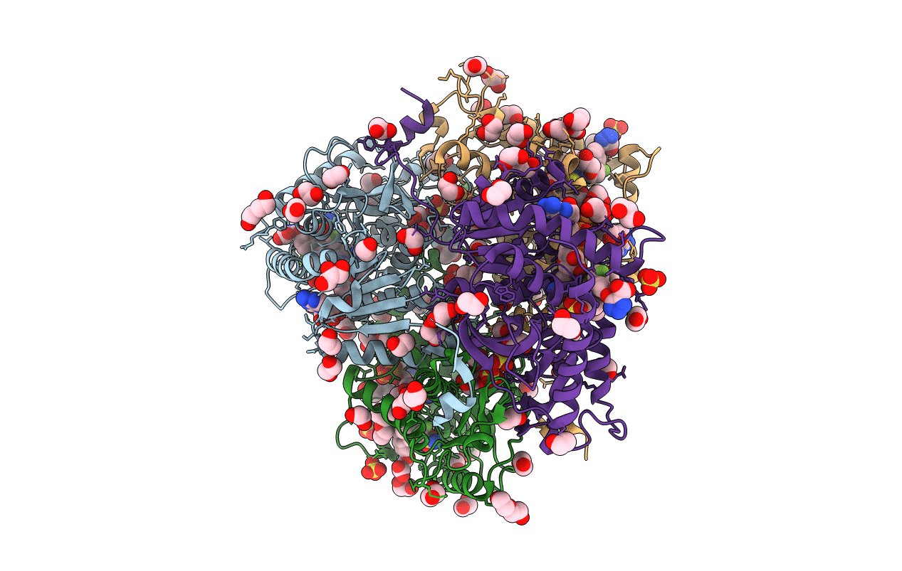 |
Organism: Homo sapiens
Method: X-RAY DIFFRACTION Resolution:2.50 Å Release Date: 2021-07-14 Classification: OXIDOREDUCTASE Ligands: NAI, SO4, EDO, GOL, PEG, YOJ |
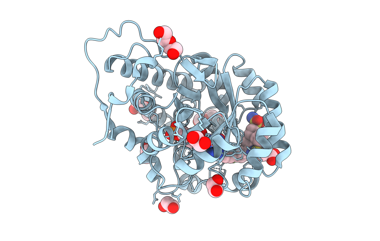 |
Organism: Homo sapiens
Method: X-RAY DIFFRACTION Resolution:2.07 Å Release Date: 2021-07-14 Classification: OXIDOREDUCTASE Ligands: FMN, YOJ, EDO, PEG |
 |
Organism: Yersinia pestis
Method: X-RAY DIFFRACTION Resolution:2.17 Å Release Date: 2019-09-11 Classification: TRANSFERASE Ligands: EDO |
 |
Organism: Legionella pneumophila subsp. pneumophila str. philadelphia 1
Method: X-RAY DIFFRACTION Resolution:2.60 Å Release Date: 2018-09-19 Classification: HYDROLASE Ligands: ZN |
 |
Crystal Structure Of Legionella Pneumophila Aminopeptidase A In Complex With Glutamic Acid
Organism: Legionella pneumophila subsp. pneumophila str. philadelphia 1
Method: X-RAY DIFFRACTION Resolution:1.86 Å Release Date: 2018-09-19 Classification: HYDROLASE Ligands: GLU |
 |
Crystal Structure Of Legionella Pneumophila Aminopeptidase A In Complex With Aspartic Acid
Organism: Legionella pneumophila subsp. pneumophila str. philadelphia 1
Method: X-RAY DIFFRACTION Resolution:2.00 Å Release Date: 2018-09-19 Classification: HYDROLASE Ligands: ASP |
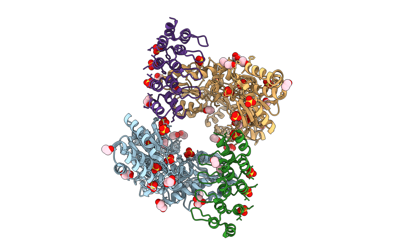 |
Crystal Structure Of The Human Dual Specificity Phosphatase 1 Catalytic Domain (C258S) As A Maltose Binding Protein Fusion In Complex With The Designed Ar Protein Off7
Organism: Escherichia coli (strain k12), Homo sapiens, Synthetic construct
Method: X-RAY DIFFRACTION Resolution:2.35 Å Release Date: 2018-09-19 Classification: HYDROLASE Ligands: GOL, SO4, EOH |
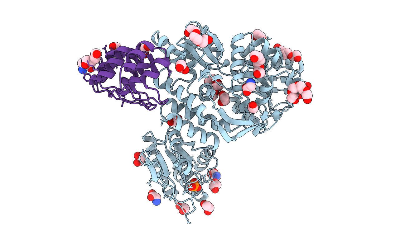 |
Crystal Structure Of The Human Dual Specificity 1 Catalytic Domain (C258S) As A Maltose Binding Protein Fusion In Complex With The Designed Ar Protein Mbp3_16
Organism: Escherichia coli (strain k12), Homo sapiens, Synthetic construct
Method: X-RAY DIFFRACTION Resolution:2.23 Å Release Date: 2018-09-19 Classification: HYDROLASE Ligands: PEG, PO4, GLY, PGE, EDO, PG4, DAL |
 |
Crystal Structure Of The Human Dual Specificity Phosphatase 1 Catalytic Domain (C258S) As A Maltose Binding Protein Fusion (Maltose Bound Form) In Complex With The Designed Ar Protein Mbp3_16
Organism: Escherichia coli (strain k12), Homo sapiens, Synthetic construct
Method: X-RAY DIFFRACTION Resolution:2.55 Å Release Date: 2018-09-19 Classification: HYDROLASE Ligands: PEG, PO4, EDO |
 |
Crystal Structure Of Human Dual Specificity Phosphatase 1 Catalytic Domain (C258S) As A Maltose Binding Protein Fusion In Complex With The Monobody Ysx1
Organism: Escherichia coli (strain k12), Homo sapiens, Synthetic construct
Method: X-RAY DIFFRACTION Resolution:2.49 Å Release Date: 2017-11-01 Classification: HYDROLASE Ligands: SO4, GOL |
 |
Crystal Structure Of Met260Ala Mutant Of E. Coli Aminopeptidase N In Complex With L-2,3-Diaminopropionic Acid
Organism: Escherichia coli k-12
Method: X-RAY DIFFRACTION Resolution:2.00 Å Release Date: 2016-03-02 Classification: HYDROLASE Ligands: ZN, DPP, NA, GOL, MLI |
 |
Crystal Structure Of Met260Ala Mutant Of E. Coli Aminopeptidase N In Complex With L-Alanine
Organism: Escherichia coli (strain k12)
Method: X-RAY DIFFRACTION Resolution:2.91 Å Release Date: 2016-03-02 Classification: HYDROLASE Ligands: ZN, ALA, NA, GOL, MLI |
 |
Crystal Structure Of Met260Ala Mutant Of E. Coli Aminopeptidase N In Complex With L-Glutamate
Organism: Escherichia coli k-12
Method: X-RAY DIFFRACTION Resolution:2.20 Å Release Date: 2016-03-02 Classification: HYDROLASE Ligands: ZN, GLU, NA, MLI |
 |
Crystal Structure Of Met260Ala Mutant Of E. Coli Aminopeptidase N In Complex With L-Leucine
Organism: Escherichia coli k-12
Method: X-RAY DIFFRACTION Resolution:2.11 Å Release Date: 2016-03-02 Classification: HYDROLASE Ligands: ZN, LEU, NA, GOL, MLI |
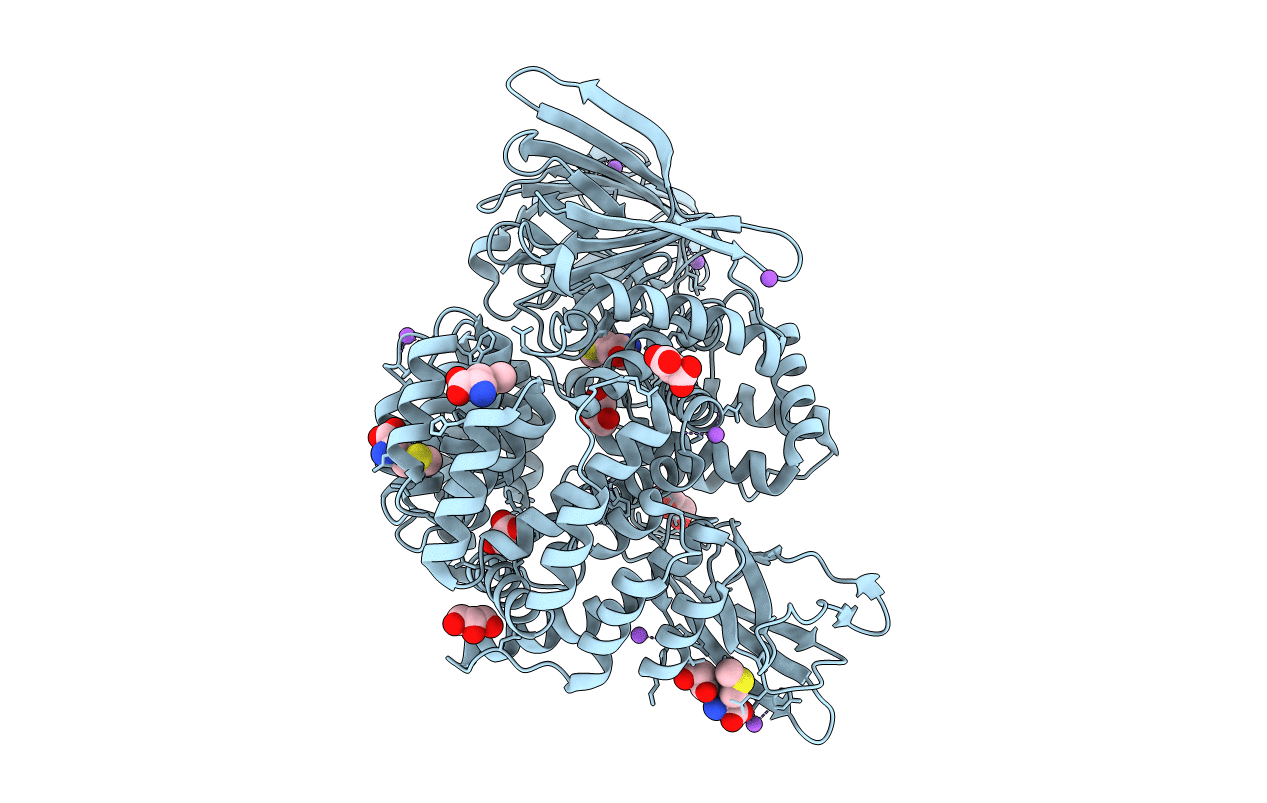 |
Crystal Structure Of Met260Ala Mutant Of E. Coli Aminopeptidase N In Complex With L-Methionine
Organism: Escherichia coli k-12
Method: X-RAY DIFFRACTION Resolution:1.99 Å Release Date: 2016-03-02 Classification: HYDROLASE Ligands: ZN, MET, NA, GOL, MLI |
 |
Crystal Structure Of E. Coli Aminopeptidase N In Complex With L-2,3-Diaminopropionic Acid
Organism: Escherichia coli k-12
Method: X-RAY DIFFRACTION Resolution:2.22 Å Release Date: 2016-03-02 Classification: HYDROLASE Ligands: ZN, DPP, NA, GOL, MLI |
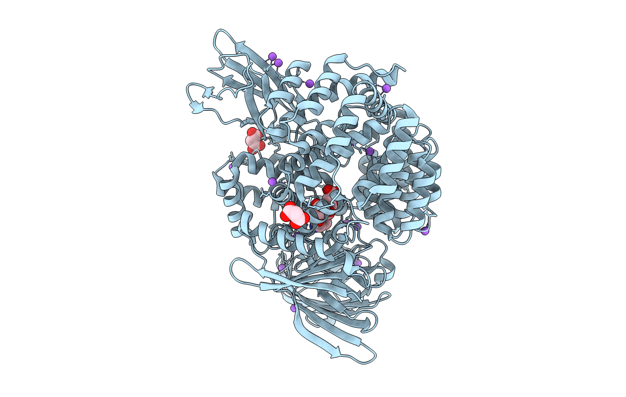 |
Organism: Escherichia coli k-12
Method: X-RAY DIFFRACTION Resolution:1.89 Å Release Date: 2016-03-02 Classification: HYDROLASE Ligands: ZN, ALA, NA, MLI |
 |
Organism: Escherichia coli k-12
Method: X-RAY DIFFRACTION Resolution:2.80 Å Release Date: 2016-03-02 Classification: HYDROLASE Ligands: ZN, BAL, NA, MLI |

