
Search Count: 135
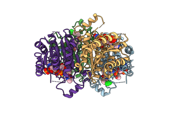 |
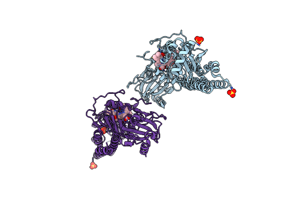
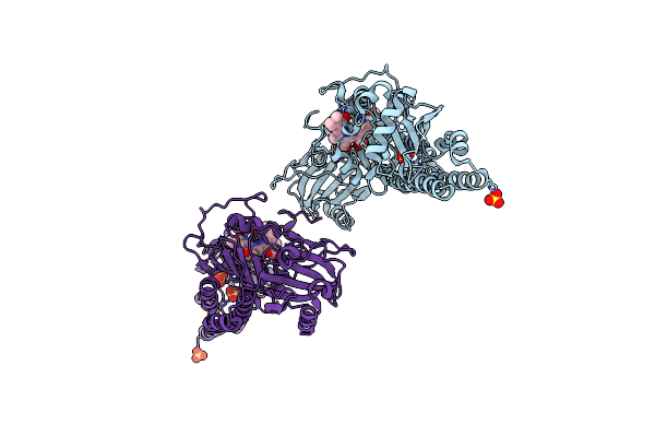
Organism: Trypanosoma brucei brucei
Method: X-RAY DIFFRACTION Release Date: 2025-10-22 Classification: OXIDOREDUCTASE Ligands: NDP, A1IXO, EDO |
 |
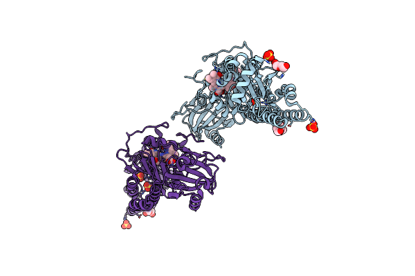
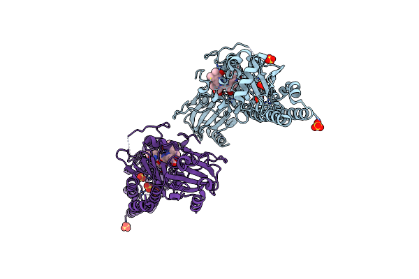
Organism: Trypanosoma brucei brucei
Method: X-RAY DIFFRACTION Release Date: 2025-10-22 Classification: OXIDOREDUCTASE Ligands: NDP, A1IXP, EDO |
 |
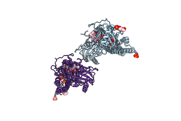
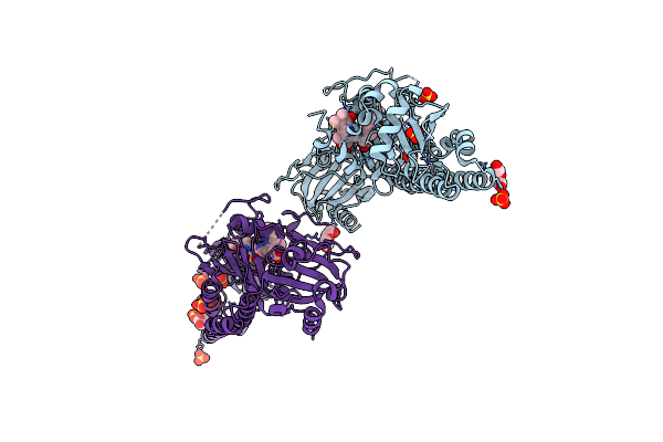
Organism: Trypanosoma brucei brucei
Method: X-RAY DIFFRACTION Release Date: 2025-10-22 Classification: OXIDOREDUCTASE Ligands: NDP, A1IXQ, ACT, EDO, CL |
 |
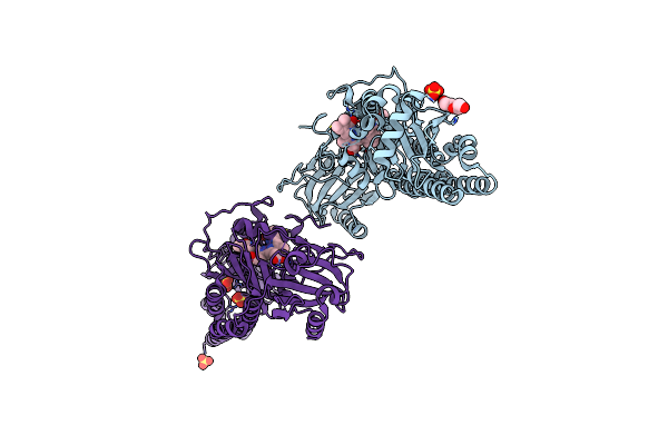
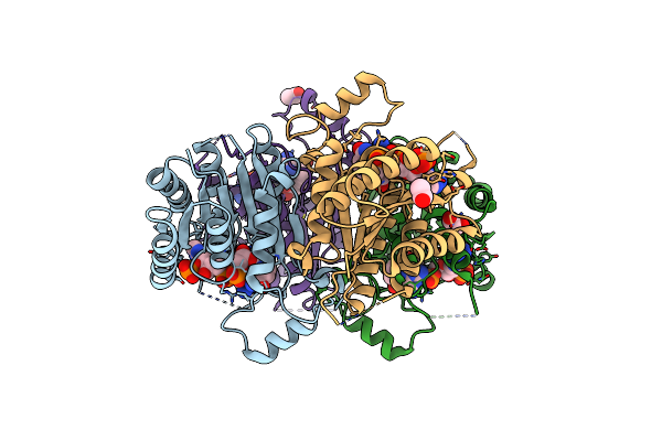
Organism: Trypanosoma brucei brucei
Method: X-RAY DIFFRACTION Release Date: 2025-10-22 Classification: OXIDOREDUCTASE Ligands: NDP, A1IXS, ACT, EDO |
 |
Organism: Trypanosoma brucei brucei
Method: X-RAY DIFFRACTION Release Date: 2025-10-22 Classification: OXIDOREDUCTASE Ligands: NDP, A1IXR, EDO |
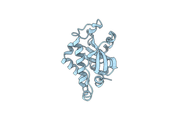 |
Crystal Structure Of The Tin2-Fold Effector Protein Tue1 From Thecaphora Thlaspeos
Organism: Thecaphora thlaspeos
Method: X-RAY DIFFRACTION Release Date: 2025-06-25 Classification: NUCLEAR PROTEIN |
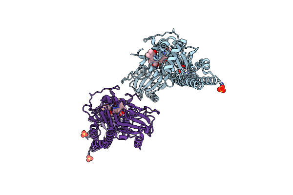 |
Serial Femtosecond X-Ray Structure Of A Fluorescence Optimized Bathy Phytochrome Pairfp2 Derived From Wild-Type Agp2 In Its Pfr State (I0A).
Organism: Agrobacterium fabrum str. c58
Method: X-RAY DIFFRACTION Release Date: 2025-05-14 Classification: SIGNALING PROTEIN Ligands: EL5, SO4, CL, EDO |
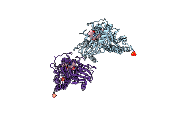 |
Serial Femtosecond X-Ray Structure Of A Fluorescence Optimized Bathy Phytochrome Pairfp2 Derived From Wild-Type Agp2 In Its Pfr State (I0B).
Organism: Agrobacterium fabrum str. c58
Method: X-RAY DIFFRACTION Release Date: 2025-05-14 Classification: SIGNALING PROTEIN Ligands: EL5, SO4 |
 |
Serial Femtosecond X-Ray Structure Of A Fluorescence Optimized Bathy Phytochrome Pairfp2 Derived From Wild-Type Agp2 In I1 Intermediate State.
Organism: Agrobacterium fabrum str. c58
Method: X-RAY DIFFRACTION Release Date: 2025-05-14 Classification: SIGNALING PROTEIN Ligands: EL5, SO4 |
 |
Serial Femtosecond X-Ray Structure Of A Fluorescence Optimized Bathy Phytochrome Pairfp2 Derived From Wild-Type Agp2 In I2 Intermediate State.
Organism: Agrobacterium fabrum str. c58
Method: X-RAY DIFFRACTION Release Date: 2025-05-14 Classification: SIGNALING PROTEIN Ligands: EL5, SO4, PGE, PEG, CL |
 |
Serial Femtosecond X-Ray Structure Of A Fluorescence Optimized Bathy Phytochrome Pairfp2 Derived From Wild-Type Agp2 In I3 Intermediate State.
Organism: Agrobacterium fabrum str. c58
Method: X-RAY DIFFRACTION Release Date: 2025-05-14 Classification: SIGNALING PROTEIN Ligands: EL5, SO4, GOL, PEG |
 |
Serial Femtosecond X-Ray Structure Of A Fluorescence Optimized Bathy Phytochrome Pairfp2 Derived From Wild-Type Agp2 In I4 Intermediate State.
Organism: Agrobacterium fabrum str. c58
Method: X-RAY DIFFRACTION Release Date: 2025-05-14 Classification: SIGNALING PROTEIN Ligands: EL5, SO4, CL, PEG |
 |
Serial Femtosecond X-Ray Structure Of A Fluorescence Optimized Bathy Phytochrome Pairfp2 Derived From Wild-Type Agp2 In I5 Intermediate State.
Organism: Agrobacterium fabrum str. c58
Method: X-RAY DIFFRACTION Release Date: 2025-05-14 Classification: SIGNALING PROTEIN Ligands: EL5, SO4, CL |
 |
Serial Femtosecond X-Ray Structure Of A Fluorescence Optimized Bathy Phytochrome Pairfp2 Derived From Wild-Type Agp2 In I6 Intermediate State.
Organism: Agrobacterium fabrum str. c58
Method: X-RAY DIFFRACTION Release Date: 2025-05-14 Classification: SIGNALING PROTEIN Ligands: EL5, SO4, CL |
 |
Serial Femtosecond X-Ray Structure Of A Fluorescence Optimized Bathy Phytochrome Pairfp2 Derived From Wild-Type Agp2 In I7 Intermediate State.
Organism: Agrobacterium fabrum str. c58
Method: X-RAY DIFFRACTION Release Date: 2025-05-14 Classification: SIGNALING PROTEIN Ligands: EL5, SO4, PEG |
 |
Crystal Structure Of Human Carbonic Anhydrase Ii In-Complex With 4-Acetylphenylboronic Acid At 2.6 A Resolution
Organism: Homo sapiens
Method: X-RAY DIFFRACTION Resolution:2.60 Å Release Date: 2024-02-28 Classification: LYASE Ligands: ZN, GOL, SO4, OY7 |
 |
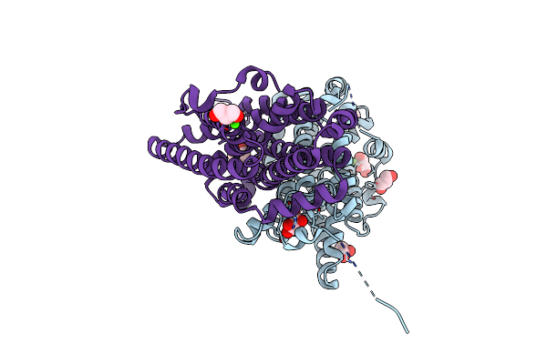
Trypanosoma Brucei Pteridine Reductase 1 (Tbptr1) In Complex With 2,4,6 Triamminopyrimidine (Tap)
Organism: Trypanosoma brucei brucei
Method: X-RAY DIFFRACTION Resolution:1.48 Å Release Date: 2023-12-13 Classification: OXIDOREDUCTASE Ligands: 3AY, NDP, EDO, ACT |
 |
Ribonucleotide Reductase Class Ie R2 From Mesoplasma Florum, Catalytically Active Radical State Solved By Xfel
Organism: Mesoplasma florum l1
Method: X-RAY DIFFRACTION Release Date: 2023-11-01 Classification: OXIDOREDUCTASE |
 |
Ribonucleotide Reductase Class Ie R2 From Mesoplasma Florum, Radical-Lost Ground State
Organism: Mesoplasma florum l1
Method: X-RAY DIFFRACTION Release Date: 2023-11-01 Classification: OXIDOREDUCTASE Ligands: CA, GOL |
 |
Xfel Structure Of Class Ib Ribonucleotide Reductase Dimanganese(Ii) Nrdf In Complex With Oxidized Nrdi From Bacillus Cereus
Organism: Bacillus cereus atcc 14579
Method: X-RAY DIFFRACTION Resolution:2.00 Å Release Date: 2022-09-21 Classification: OXIDOREDUCTASE Ligands: MN, UNX, FMN |