
Search Count: 19
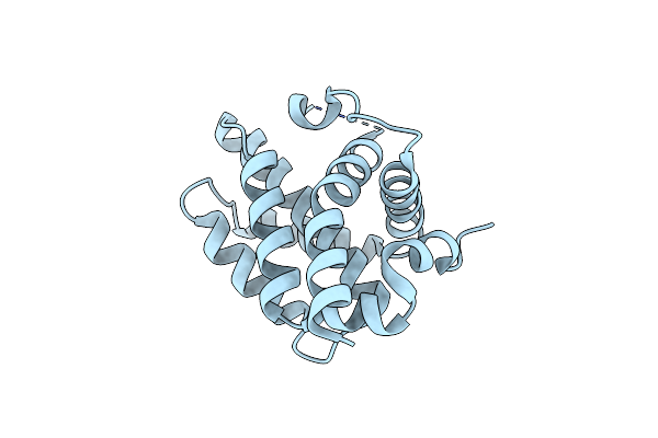 |
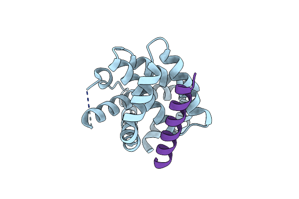
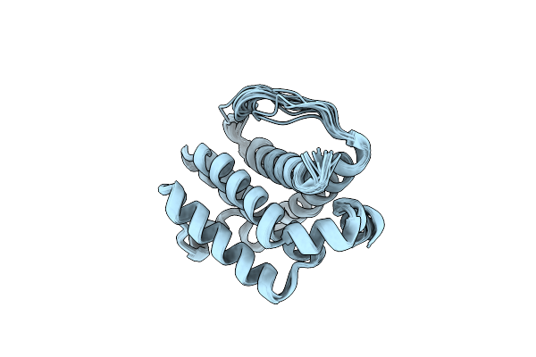
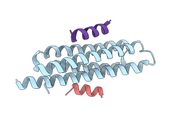
Structural Basis Of Bak Sequestration By Mcl-1 And Consequences For Apoptosis Initiation
Organism: Homo sapiens
Method: X-RAY DIFFRACTION Resolution:1.49 Å Release Date: 2025-06-04 Classification: APOPTOSIS |
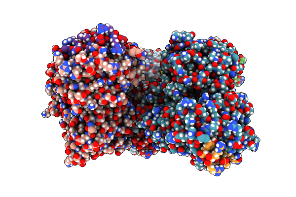 |
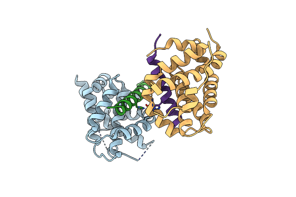
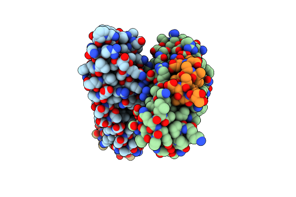
Structural Basis Of Bak Sequestration By Mcl-1 And Consequences For Apoptosis Initiation
Organism: Synthetic construct, Homo sapiens
Method: X-RAY DIFFRACTION Resolution:1.70 Å Release Date: 2025-06-04 Classification: APOPTOSIS |
 |
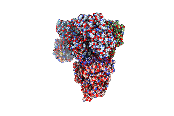
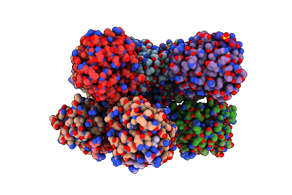
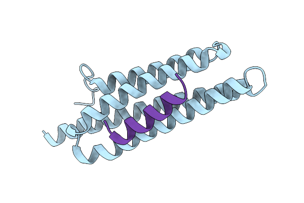
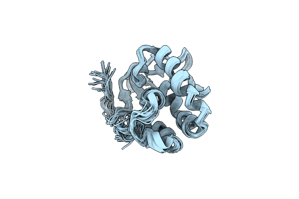
Structural Basis Of Bak Sequestration By Mcl-1 And Consequences For Apoptosis Initiation
Organism: Homo sapiens
Method: ELECTRON MICROSCOPY Release Date: 2025-06-04 Classification: APOPTOSIS |
 |
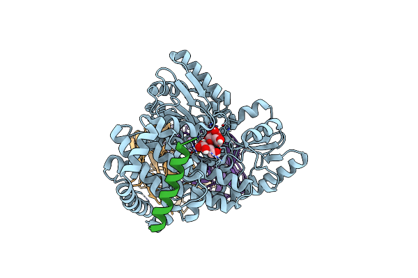
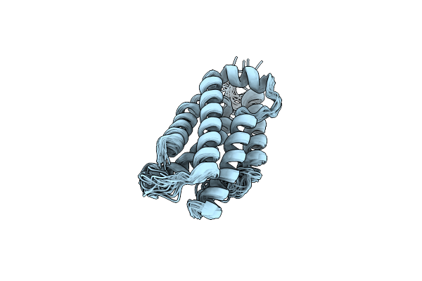
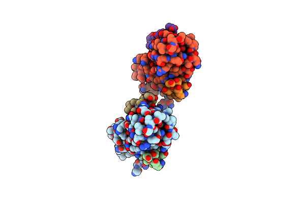
Structural Basis Of Bak Sequestration By Mcl-1 And Consequences For Apoptosis Initiation
Organism: Synthetic construct, Homo sapiens
Method: X-RAY DIFFRACTION Resolution:1.89 Å Release Date: 2025-06-04 Classification: APOPTOSIS |
 |
Organism: Homo sapiens
Method: X-RAY DIFFRACTION Resolution:1.50 Å Release Date: 2022-01-12 Classification: APOPTOSIS |
 |
Organism: Homo sapiens
Method: X-RAY DIFFRACTION Resolution:1.85 Å Release Date: 2022-01-12 Classification: APOPTOSIS Ligands: CU |
 |
Organism: Homo sapiens
Method: X-RAY DIFFRACTION Resolution:3.06 Å Release Date: 2022-01-12 Classification: APOPTOSIS Ligands: CU, SO4 |
 |
Direct Activation Of The Executioner Domain Of Mlkl By A Select Repertoire Of Inositol Phosphates
Organism: Homo sapiens
Method: SOLUTION NMR Release Date: 2019-05-15 Classification: LIPID BINDING PROTEIN |
 |
 |
Organism: Mus musculus
Method: X-RAY DIFFRACTION Resolution:2.00 Å Release Date: 2017-09-06 Classification: CELL ADHESION |
 |
Organism: Homo sapiens
Method: X-RAY DIFFRACTION Resolution:1.79 Å Release Date: 2015-12-30 Classification: CELL ADHESION |
 |
Fusion Of Pyk2-Fat Domain With Leupaxin Ld1 Motif, Complexed With Leupaxin Ld4 Peptide
Organism: Homo sapiens
Method: X-RAY DIFFRACTION Resolution:2.01 Å Release Date: 2015-12-30 Classification: CELL ADHESION |
 |
Organism: Homo sapiens
Method: X-RAY DIFFRACTION Resolution:2.50 Å Release Date: 2015-12-23 Classification: CELL ADHESION |
 |
Organism: Homo sapiens, Gallus gallus
Method: X-RAY DIFFRACTION Resolution:3.51 Å Release Date: 2014-09-17 Classification: CELL ADHESION |
 |
Organism: Drosophila melanogaster
Method: SOLUTION NMR Release Date: 2014-03-05 Classification: ONCOPROTEIN |
 |
Structural Basis For The Interaction Of Pyk2 Pat Domain With Paxillin Ld Motifs
Organism: Homo sapiens
Method: X-RAY DIFFRACTION Resolution:3.10 Å Release Date: 2012-10-24 Classification: TRANSFERASE/signaling protein |
 |
Structural And Mechanistic Insights Into The Interaction Between Pat Pyk2 And Paxillin Ld Motif
|
 |
Organism: Gallus gallus
Method: SOLUTION NMR Release Date: 2008-04-29 Classification: STRUCTURAL PROTEIN |
 |
The Paxillin-Binding Domain (Pbd) Of G Protein Coupled Receptor (Gpcr)-Kinase (Grk) Interacting Protein 1 (Git1)
Organism: Rattus norvegicus
Method: SOLUTION NMR Release Date: 2008-04-29 Classification: CELL ADHESION, SIGNALING PROTEIN |