
Search Count: 5
 |
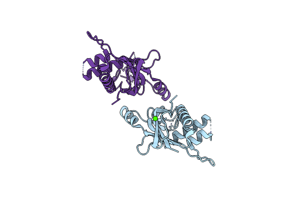
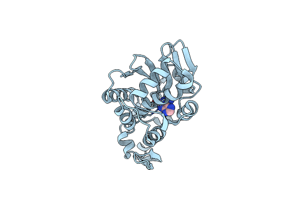
Crystal Structure Of The C-Terminal Periplasmic Domain Of Eceptc From Escherichia Coli- Complex With Zn
Organism: Escherichia coli
Method: X-RAY DIFFRACTION Resolution:2.10 Å Release Date: 2019-06-05 Classification: TRANSFERASE Ligands: ZN |
 |
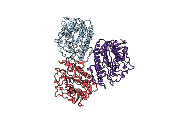
Structural Insights Into The Crispr-Cas-Associated Ribonuclease Activity Of Staphylococcus Epidermidis Csm6
Organism: Staphylococcus epidermidis (strain atcc 35984 / rp62a)
Method: X-RAY DIFFRACTION Resolution:2.01 Å Release Date: 2018-10-17 Classification: HYDROLASE |
 |
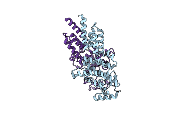
Structural Insights Into The Crispr-Cas-Associated Ribonuclease Activity Of Staphylococcus Epidermidis Csm3
Organism: Staphylococcus epidermidis rp62a
Method: X-RAY DIFFRACTION Resolution:2.26 Å Release Date: 2018-10-17 Classification: HYDROLASE Ligands: CA |
 |
Crystal Structure Of Mycobacterium Tuberculosis Shikimate Kinase In Complex With Shikimate And Amppcp At 2.85 Angstrom Resolution
Organism: Mycobacterium tuberculosis
Method: X-RAY DIFFRACTION Resolution:2.90 Å Release Date: 2006-07-11 Classification: SIGANLING PROTEIN,TRANSFERASE Ligands: ACP, SKM |
 |
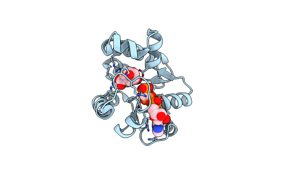
Crystal Structure Of The Complex Of Trichosanthin With Adenine, Obtained From Trichosanthin Complexed With The Dinucleotide Apg
Organism: Trichosanthes kirilowii
Method: X-RAY DIFFRACTION Resolution:1.86 Å Release Date: 2000-04-24 Classification: HYDROLASE Ligands: ADE |