Planned Maintenance: Some services may turn out to be unavailable from 15th January, 2026 to 16th January, 2026. We apologize for the inconvenience!

Planned Maintenance: Some services may turn out to be unavailable from 15th January, 2026 to 16th January, 2026. We apologize for the inconvenience!
 |
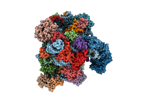
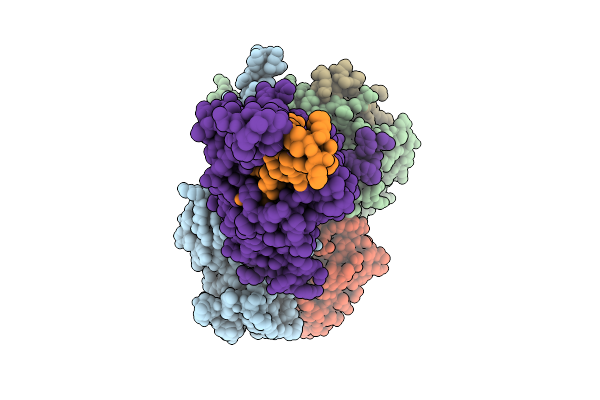
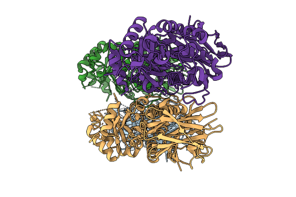
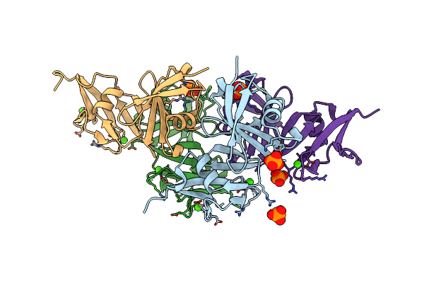
Organism: Goslarvirus
Method: ELECTRON MICROSCOPY Release Date: 2025-12-24 Classification: VIRAL PROTEIN Ligands: G2P |
 |
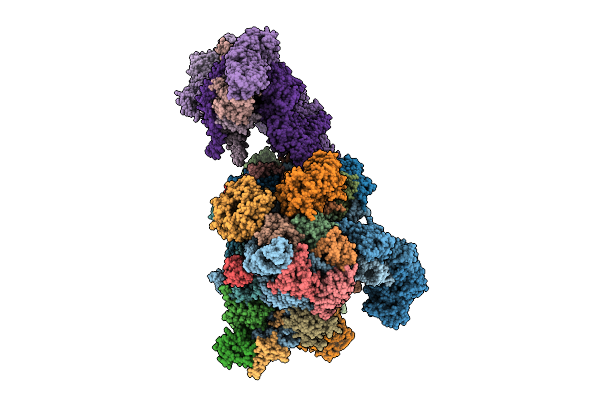
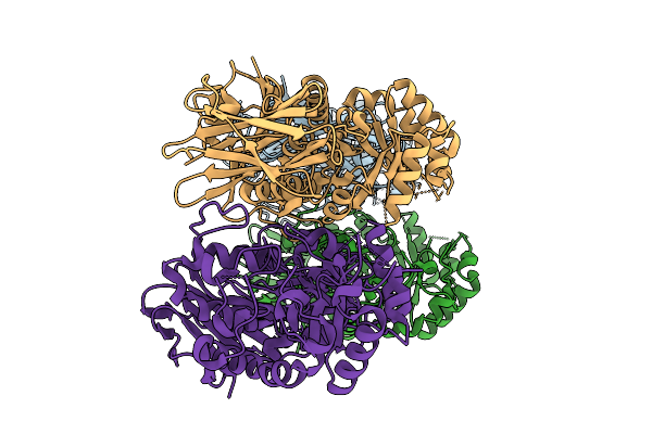
Organism: Goslarvirus
Method: ELECTRON MICROSCOPY Release Date: 2025-12-24 Classification: VIRAL PROTEIN Ligands: G2P |
 |
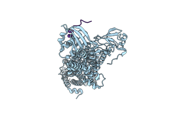
Organism: Oryctolagus cuniculus
Method: ELECTRON MICROSCOPY Release Date: 2025-12-10 Classification: TRANSLATION Ligands: SF4 |
 |
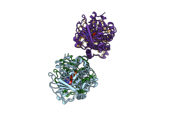
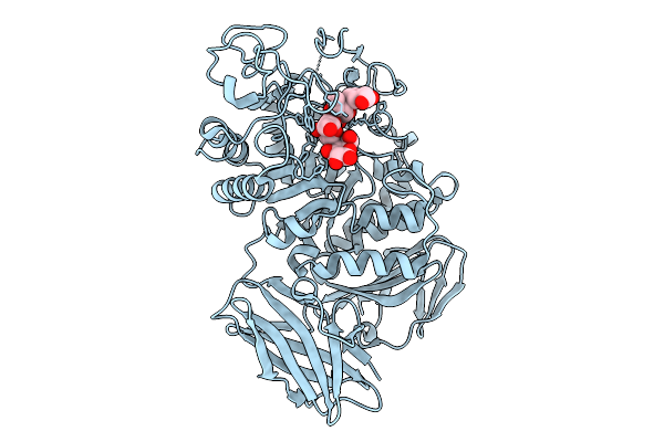
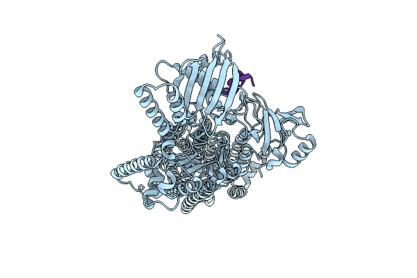
Late-Stage 48S Initiation Complex With Eif3 (Ls48S-Eif3 Ic) Guided By The Trans-Rna
Organism: Oryctolagus cuniculus
Method: ELECTRON MICROSCOPY Release Date: 2025-12-10 Classification: TRANSLATION Ligands: SF4 |
 |
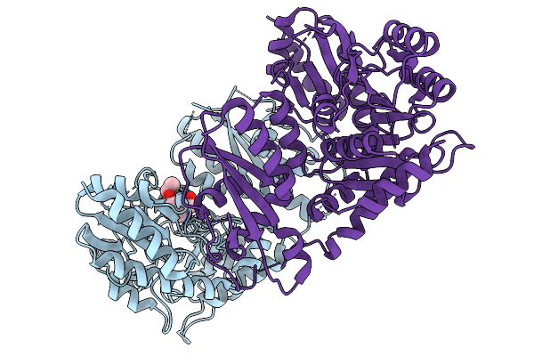
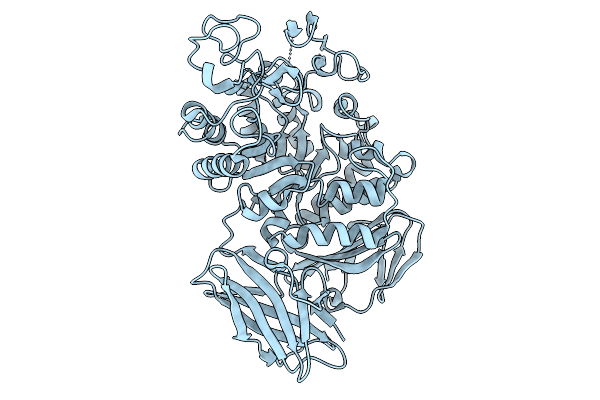
Organism: Penicillium citrinum
Method: X-RAY DIFFRACTION Release Date: 2025-11-26 Classification: LYASE Ligands: PO4, CL |
 |
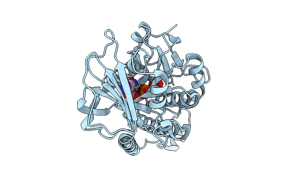
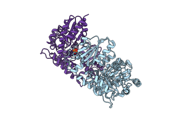
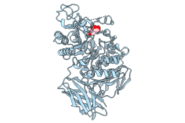
Gamma-Lyase Cndf In Complex With Pyridoxal 5'-Phosphate, L-Homoserine And Ethyl Acetoacetate
Organism: Penicillium citrinum
Method: X-RAY DIFFRACTION Release Date: 2025-11-19 Classification: LYASE Ligands: HSE, EAC |
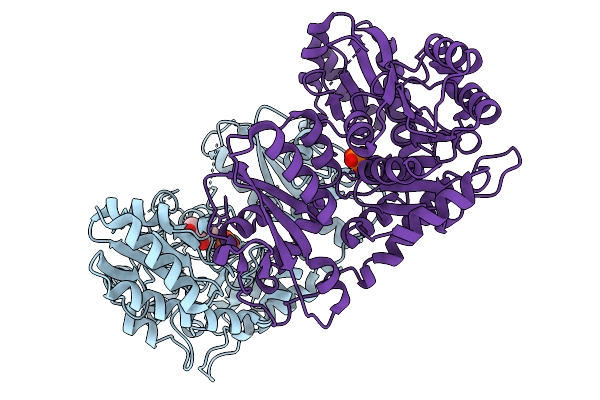 |
Organism: Penicillium citrinum
Method: X-RAY DIFFRACTION Release Date: 2025-11-19 Classification: LYASE Ligands: PO4, EAC |
 |
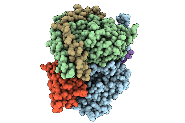
Organism: Synechococcus elongatus, Synechococcus elongatus pcc 7942 = fachb-805, Synthetic construct
Method: ELECTRON MICROSCOPY Release Date: 2025-10-15 Classification: DNA BINDING PROTEIN/DNA Ligands: MG, ZN |
 |
Cryo-Em Structure Of Rpaa Bound To Pkaibc Dna And The Ctd Of The Alpha Subunit Of Rnap From The Cyanobacterium Synechococcus Elongatus
Organism: Synechococcus elongatus, Synechococcus elongatus pcc 7942 = fachb-805, Synthetic construct
Method: ELECTRON MICROSCOPY Release Date: 2025-10-15 Classification: DNA BINDING PROTEIN/DNA |
 |
Cryoem Structure Of Rpaa Bound To Pkaibc Dna And Syn7942 Rnap-Siga Holoenzyme
Organism: Synechococcus elongatus, Synechococcus elongatus pcc 7942 = fachb-805, Synthetic construct
Method: ELECTRON MICROSCOPY Release Date: 2025-10-15 Classification: DNA BINDING PROTEIN/DNA Ligands: MG, ZN |
 |
Organism: Homo sapiens, Mus musculus
Method: ELECTRON MICROSCOPY Release Date: 2025-09-03 Classification: MEMBRANE PROTEIN |
 |
Organism: Homo sapiens, Mus musculus
Method: ELECTRON MICROSCOPY Release Date: 2025-09-03 Classification: MEMBRANE PROTEIN |
 |
Apg Mutant Enzyme D448N Of Acarbose Hydrolase From Human Gut Flora K. Grimontii Td1,Complex With Acarbose
Organism: Klebsiella grimontii
Method: X-RAY DIFFRACTION Release Date: 2025-09-03 Classification: HYDROLASE |
 |
Apg Mutant Enzyme D448N Of The Human Gut Flora K. Grimontii Td1 Acarbose Hydrolase
Organism: Klebsiella grimontii
Method: X-RAY DIFFRACTION Release Date: 2025-09-03 Classification: HYDROLASE |
 |
Structure Of A Truncated Loopa Mutant From The Human Gut Flora K. Grimontii Apg In Complex With Glucose
Organism: Klebsiella grimontii
Method: X-RAY DIFFRACTION Release Date: 2025-09-03 Classification: HYDROLASE Ligands: GLC |
 |
Structure Of A Truncated Loopb Mutant From The Human Gut Flora K. Grimontii Apg
Organism: Klebsiella grimontii
Method: X-RAY DIFFRACTION Release Date: 2025-09-03 Classification: HYDROLASE |
 |
Structure Of Truncated Loopa And Loopb Mutants From The Human Gut Flora K. Grimontii Apg
Organism: Klebsiella grimontii
Method: X-RAY DIFFRACTION Release Date: 2025-09-03 Classification: HYDROLASE |
 |
Cryo-Em Structure Of The Pma1 With Ordered N-Terminal Extension In The Activated State
Organism: Saccharomyces cerevisiae (strain atcc 204508 / s288c)
Method: ELECTRON MICROSCOPY Release Date: 2025-06-04 Classification: PROTON TRANSPORT |
 |
Cryo-Em Structure Of The Pma1 With Ordered N-Terminal Extension In The Autoinhibited State
Organism: Saccharomyces cerevisiae (strain atcc 204508 / s288c)
Method: ELECTRON MICROSCOPY Release Date: 2025-06-04 Classification: PROTON TRANSPORT |
 |
Organism: Methylobacterium brachiatum
Method: X-RAY DIFFRACTION Release Date: 2025-04-09 Classification: PROTEIN BINDING Ligands: CA, PO4 |