
Search Count: 12
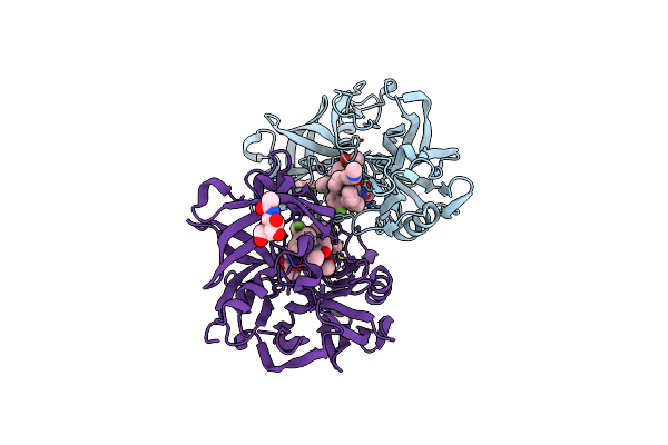 |
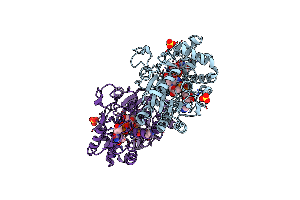
Crystal Structure Of Human Renin In Complex With A Biphenylpipderidinylcarbinol
Organism: Homo sapiens
Method: X-RAY DIFFRACTION Resolution:2.60 Å Release Date: 2017-10-18 Classification: HYDROLASE/HYDROLASE inhibitor Ligands: NAG, 90D |
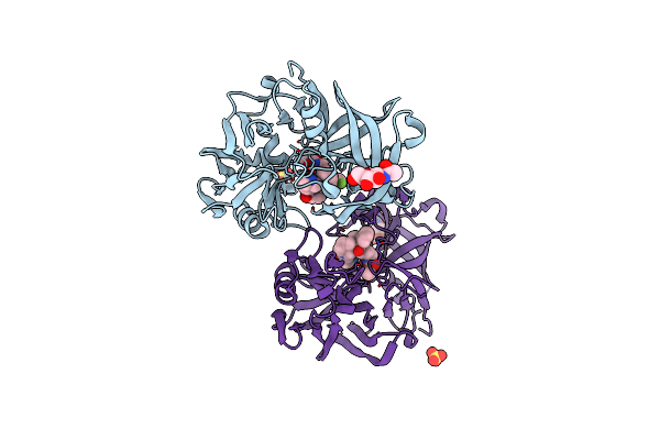 |
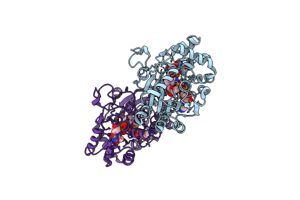
Crystal Structure Of Human Renin In Complex With A Biphenylpipderidinylcarbinol
Organism: Homo sapiens
Method: X-RAY DIFFRACTION Resolution:2.90 Å Release Date: 2017-10-18 Classification: HYDROLASE/HYDROLASE inhibitor Ligands: NAG, 9G7, SO4 |
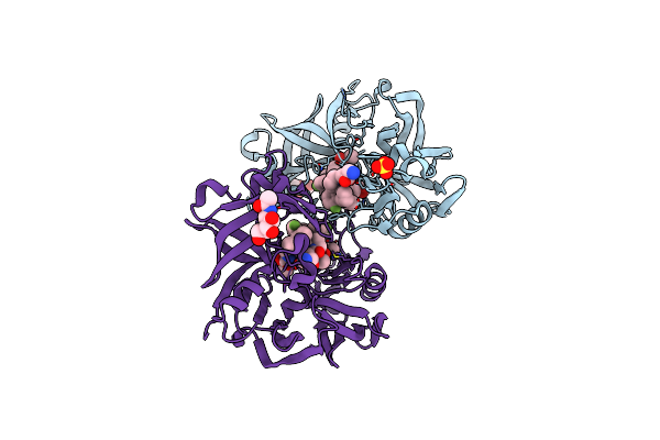 |
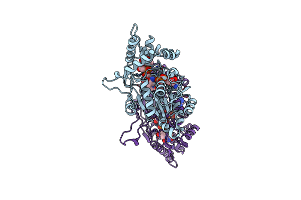
Crystal Structure Of Human Renin In Complex With A Biphenylpipderidinylcarbinol
Organism: Homo sapiens
Method: X-RAY DIFFRACTION Resolution:3.22 Å Release Date: 2017-10-18 Classification: HYDROLASE/HYDROLASE inhibitor Ligands: NAG, SO4, 9JD |
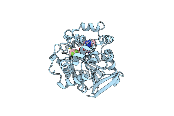 |
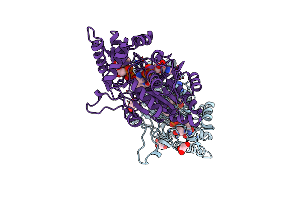
Organism: Homo sapiens
Method: X-RAY DIFFRACTION Resolution:1.96 Å Release Date: 2013-06-05 Classification: Hydrolase/hydrolase inhibitor Ligands: 1LF |
 |
Organism: Homo sapiens
Method: X-RAY DIFFRACTION Resolution:2.00 Å Release Date: 2009-09-08 Classification: HYDROLASE Ligands: ZN, CA, 077, SO4, 023, GOL |
 |
Organism: Homo sapiens
Method: X-RAY DIFFRACTION Resolution:1.70 Å Release Date: 2009-09-08 Classification: HYDROLASE Ligands: ZN, CA, 068, SO4, 023, GOL |
 |
Organism: Homo sapiens
Method: X-RAY DIFFRACTION Resolution:2.30 Å Release Date: 2009-09-08 Classification: HYDROLASE Ligands: SO4, ZN, CA, 576, 023, GOL |
 |
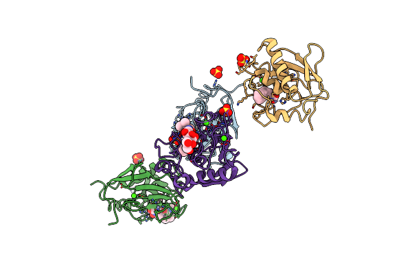
The Crystal Structure Of Dtdp-D-Glucose 4,6-Dehydratase (Rmlb) From Streptococcus Suis With Dtdp-Xylose Bound
Organism: Streptococcus suis
Method: X-RAY DIFFRACTION Resolution:1.80 Å Release Date: 2002-01-25 Classification: LYASE Ligands: SO4, NAD, TDX |
 |
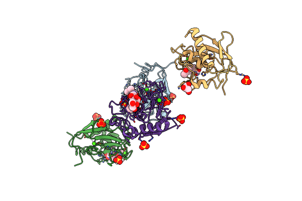
The Crystal Structure Of Dtdp-D-Glucose 4,6-Dehydratase (Rmlb) From Streptococcus Suis With Dtdp-D-Glucose Bound
Organism: Streptococcus suis
Method: X-RAY DIFFRACTION Resolution:2.20 Å Release Date: 2002-01-25 Classification: LYASE Ligands: SO4, DAU, NAD |
 |
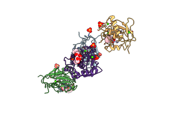
The Crystal Structure Of Dtdp-D-Glucose 4,6-Dehydratase (Rmlb) From Streptococcus Suis With Thymidine Diphosphate Bound
Organism: Streptococcus suis
Method: X-RAY DIFFRACTION Resolution:1.80 Å Release Date: 2002-01-25 Classification: LYASE Ligands: TYD, NAD |
 |
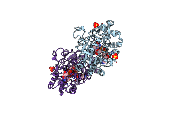
The Crystal Structure Of Dtdp-D-Glucose 4,6-Dehydratase (Rmlb) From Salmonella Enterica Serovar Typhimurium With Dtdp-D-Glucose Bound
Organism: Salmonella enterica subsp. enterica serovar typhimurium
Method: X-RAY DIFFRACTION Resolution:2.40 Å Release Date: 2002-01-25 Classification: LYASE Ligands: DAU, NAD |
 |
The Crystal Structure Of Dtdp-D-Glucose 4,6-Dehydratase (Rmlb) From Salmonella Enterica Serovar Typhimurium With Thymidine Diphosphate Bound
Organism: Salmonella enterica subsp. enterica serovar typhimurium
Method: X-RAY DIFFRACTION Resolution:1.80 Å Release Date: 2002-01-25 Classification: LYASE Ligands: TYD, NAD, GOL |