
Search Count: 26
 |
 |
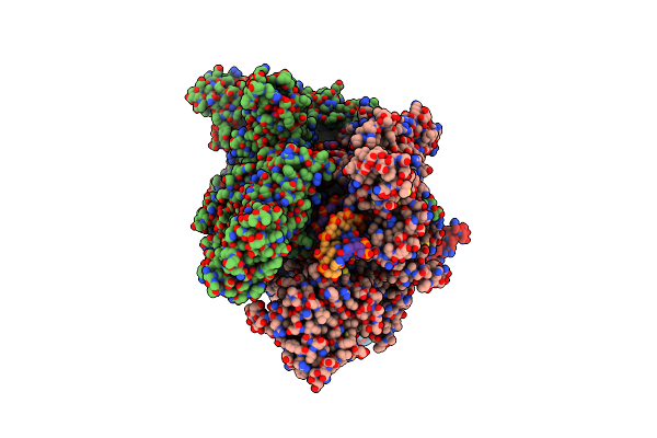
Organism: Dunaliella tertiolecta
Method: ELECTRON MICROSCOPY Release Date: 2025-06-25 Classification: PHOTOSYNTHESIS Ligands: CHL, CLA, LUT, XAT, BCR, LHG, SQD, DGD, LMG, LMU, PTY, CL0, PQN, SF4, LMK |
 |
 |
Organism: Dunaliella tertiolecta
Method: ELECTRON MICROSCOPY Release Date: 2025-06-25 Classification: PHOTOSYNTHESIS Ligands: CHL, CLA, LUT, XAT, BCR, LHG, SQD, PTY, LMG, DGD, LMU, CL0, PQN, SF4, LMK |
 |
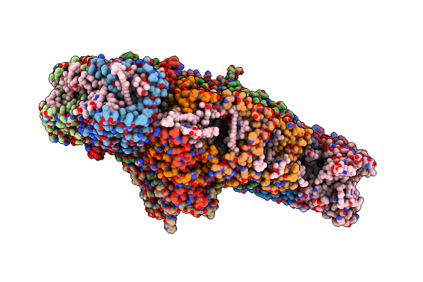
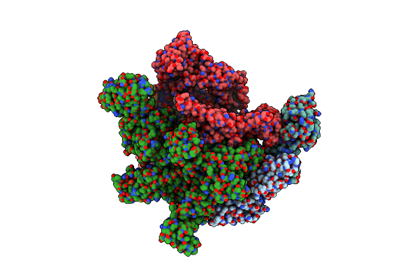
Cryo-Em Structure Of Human Rna Polymerase Ii Elongation Complex In An Intermediate Translocation State
Organism: Homo sapiens
Method: ELECTRON MICROSCOPY Resolution:2.60 Å Release Date: 2025-03-05 Classification: TRANSCRIPTION, TRANSFERASE/DNA/RNA Ligands: ZN, MG |
 |
Cryo-Em Structure Of Human Rna Polymerase Ii Elongation Complex Bound To The Recql5 Helicase In The Absence Of Nucleotide
Method: ELECTRON MICROSCOPY
Resolution:3.20 Å Release Date: 2025-03-05 Classification: TRANSCRIPTION, TRANSFERASE/DNA/RNA Ligands: ZN, MG |
 |
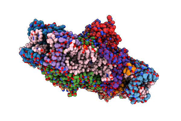
Cryo-Em Structure Of Human Rna Polymerase Ii Elongation Complex Bound To An Apo Recql5 Helicase (Recql5 Iri Module Focused-Classified)
Organism: Homo sapiens
Method: ELECTRON MICROSCOPY Resolution:2.80 Å Release Date: 2025-03-05 Classification: TRANSCRIPTION, TRANSFERASE/DNA/RNA |
 |
Cryo-Em Structure Of Human Rna Polymerase Ii Elongation Complex Bound To The Recql5 Helicase In The Presence Of Amppnp
Method: ELECTRON MICROSCOPY
Release Date: 2025-03-05 Classification: TRANSCRIPTION, TRANSFERASE/DNA/RNA Ligands: ZN, MG, ANP |
 |
Cryo-Em Structure Of Human Rna Polymerase Ii Elongation Complex Bound To The Recql5 Helicase In The Presence Of Adp
Method: ELECTRON MICROSCOPY
Release Date: 2025-03-05 Classification: TRANSCRIPTION, TRANSFERASE/DNA/RNA Ligands: ZN, MG, ADP |
 |
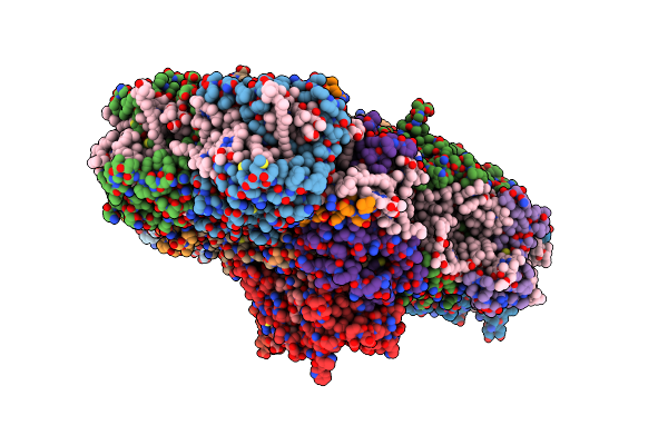
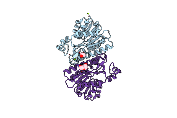
Cryo-Em Structure Of E. Coli Rna Polymerase Backtracked Elongation Complex Harboring A Terminal Mismatch
Organism: Escherichia coli k-12, Escherichia phage lambda
Method: ELECTRON MICROSCOPY Release Date: 2023-03-29 Classification: TRANSCRIPTION Ligands: ZN, MG |
 |
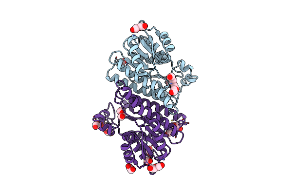
Cryo-Em Structure Of E. Coli Rna Polymerase Elongation Complex In The Transcription-Translation Complex (Rnap In An Anti-Swiveled Conformation)
Organism: Escherichia coli k-12, Escherichia phage lambda
Method: ELECTRON MICROSCOPY Release Date: 2023-03-29 Classification: TRANSCRIPTION/DNA/RNA Ligands: ZN, MG |
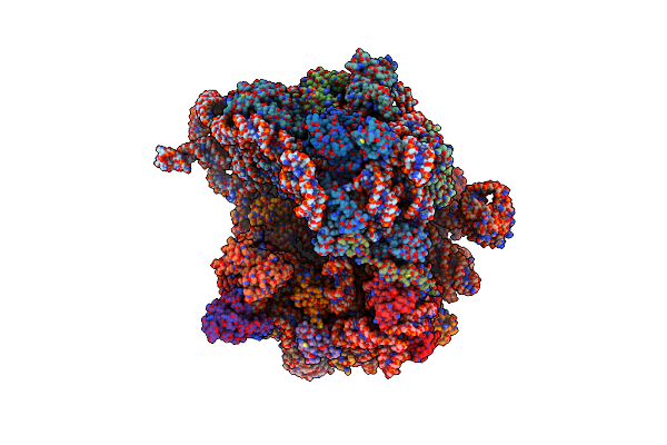 |
Cryo-Em Structure Of E. Coli 70S Ribosome Containing Mrna And Trna (In The Transcription-Translation Complex)
Organism: Escherichia phage lambda, Escherichia coli
Method: ELECTRON MICROSCOPY Release Date: 2023-03-29 Classification: TRANSLATION |
 |
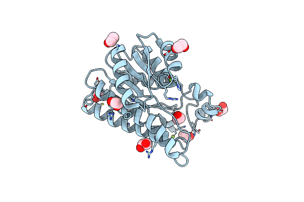
Organism: Lactobacillus brevis
Method: X-RAY DIFFRACTION Resolution:1.40 Å Release Date: 2020-10-28 Classification: OXIDOREDUCTASE Ligands: MG |
 |
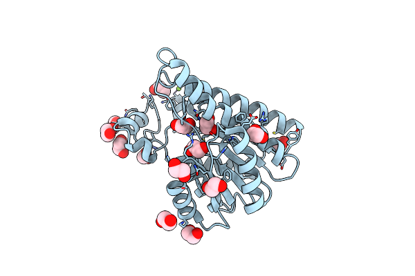
Organism: Lactobacillus brevis
Method: X-RAY DIFFRACTION Resolution:1.44 Å Release Date: 2020-02-19 Classification: OXIDOREDUCTASE Ligands: MG |
 |
Organism: Lactobacillus brevis
Method: X-RAY DIFFRACTION Resolution:1.21 Å Release Date: 2020-02-19 Classification: OXIDOREDUCTASE Ligands: EDO, PEG, PGE, MG |
 |
Organism: Lactobacillus brevis
Method: X-RAY DIFFRACTION Resolution:1.22 Å Release Date: 2020-02-19 Classification: OXIDOREDUCTASE Ligands: MG, EDO |
 |
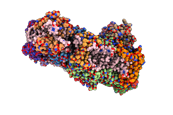
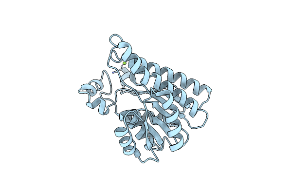
X-Ray Structure Of Lactobacillus Brevis Alcohol Dehydrogenase Mutant T102E_Q126K
Organism: Lactobacillus brevis
Method: X-RAY DIFFRACTION Resolution:1.80 Å Release Date: 2020-02-19 Classification: OXIDOREDUCTASE Ligands: EDO, MG |
 |
X-Ray Structure Of Lactobacillus Brevis Alcohol Dehydrogenase Mutant K32A_Q126K
Organism: Lactobacillus brevis
Method: X-RAY DIFFRACTION Resolution:1.40 Å Release Date: 2020-02-19 Classification: OXIDOREDUCTASE Ligands: EDO, MG, PEG |
 |
Organism: Lactobacillus brevis
Method: X-RAY DIFFRACTION Resolution:1.41 Å Release Date: 2020-02-19 Classification: OXIDOREDUCTASE Ligands: PEG, PG4, EDO, MG, CL, PGE |
 |
A Unique Supramolecular Organization Of Photosystem I In The Moss Physcomitrella Patens
Organism: Physcomitrella patens
Method: ELECTRON MICROSCOPY Resolution:11.60 Å Release Date: 2018-11-21 Classification: PHOTOSYNTHESIS |