
Search Count: 15
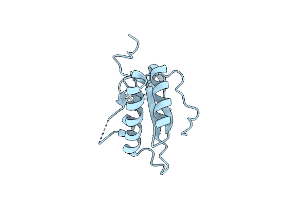 |
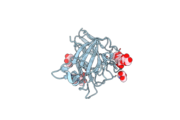
Organism: Escherichia phage ecd7
Method: X-RAY DIFFRACTION Resolution:2.49 Å Release Date: 2024-07-31 Classification: ANTIMICROBIAL PROTEIN |
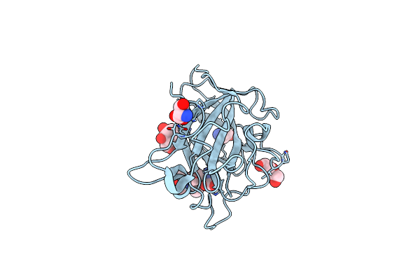 |
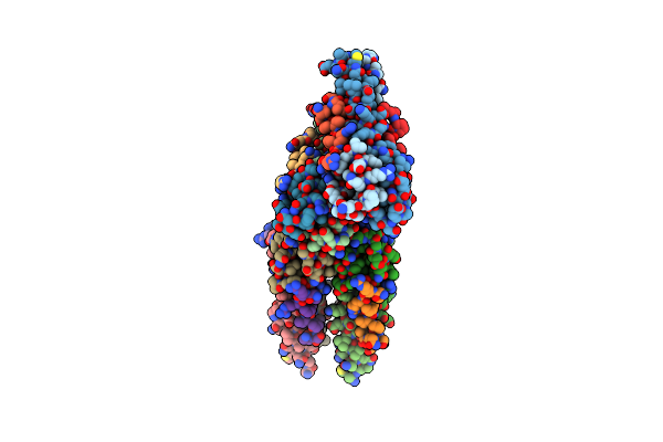
Organism: Lentinus similis
Method: X-RAY DIFFRACTION Resolution:1.60 Å Release Date: 2019-09-11 Classification: UNKNOWN FUNCTION Ligands: MAN, CL, PEG, GLY, SER |
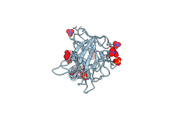 |
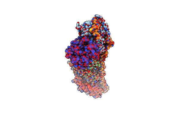
Organism: Lentinus similis
Method: X-RAY DIFFRACTION Resolution:1.60 Å Release Date: 2019-09-11 Classification: UNKNOWN FUNCTION Ligands: MAN, MPD, SO4, NO3, PGE |
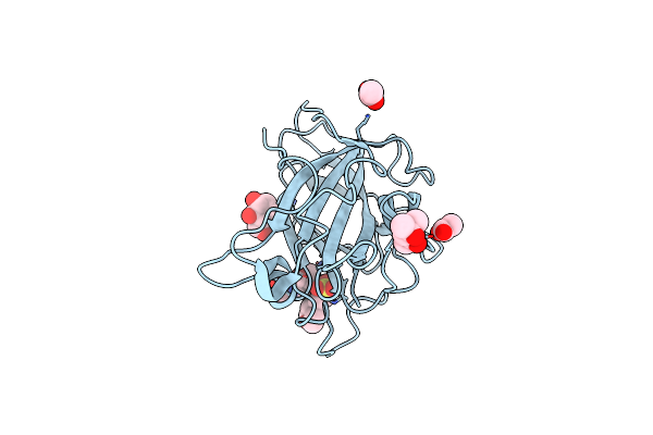 |
Organism: Lentinus similis
Method: X-RAY DIFFRACTION Resolution:1.58 Å Release Date: 2019-09-11 Classification: UNKNOWN FUNCTION Ligands: MAN, MES, MPD, ACT |
 |
Organism: Lentinus similis
Method: X-RAY DIFFRACTION Resolution:1.40 Å Release Date: 2019-09-11 Classification: UNKNOWN FUNCTION Ligands: MAN, BCN |
 |
Organism: Homo sapiens, Saccharomyces cerevisiae
Method: X-RAY DIFFRACTION Resolution:2.30 Å Release Date: 2017-06-14 Classification: microtubule binding protein |
 |
Organism: Rattus norvegicus, Gallus gallus, Bos taurus
Method: X-RAY DIFFRACTION Resolution:2.25 Å Release Date: 2016-06-29 Classification: CELL CYCLE Ligands: GTP, MG, CA, IMD, GOL, GDP, 6K9, 03S, MES, ACP |
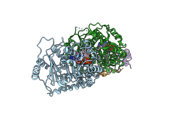 |
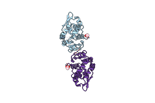
Crystal Structure Of The Sac Domain Of Cpap In A Complex With Tubulin And Darpin
Organism: Homo sapiens, Synthetic construct, Bos taurus
Method: X-RAY DIFFRACTION Resolution:2.20 Å Release Date: 2016-06-01 Classification: STRUCTURAL PROTEIN Ligands: GTP, MG, GDP, LOC |
 |
Organism: Homo sapiens
Method: X-RAY DIFFRACTION Resolution:1.75 Å Release Date: 2011-06-29 Classification: STRUCTURAL PROTEIN |
 |
Crystal Structure Of Human Eb1 In Complex With Microtubule Tip Localization Signal Peptide Of Macf
Organism: Homo sapiens
Method: X-RAY DIFFRACTION Resolution:2.50 Å Release Date: 2009-08-04 Classification: STRUCTURAL PROTEIN |
 |
Organism: Homo sapiens
Method: X-RAY DIFFRACTION Resolution:1.40 Å Release Date: 2009-03-17 Classification: PROTEIN BINDING |
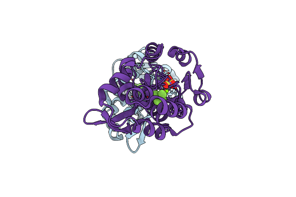 |
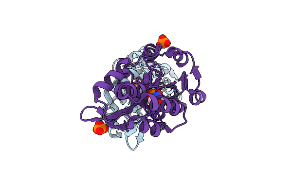
Transition State Analogue Of Phosphoserine Phosphatase (Aluminum Fluoride Complex)
Organism: Methanocaldococcus jannaschii
Method: X-RAY DIFFRACTION Resolution:1.80 Å Release Date: 2002-06-19 Classification: HYDROLASE Ligands: ALF, MG, SO4, AF3 |
 |
Organism: Methanocaldococcus jannaschii
Method: X-RAY DIFFRACTION Resolution:2.20 Å Release Date: 2002-06-19 Classification: HYDROLASE Ligands: ZN, ACY |
 |
Organism: Methanocaldococcus jannaschii
Method: X-RAY DIFFRACTION Resolution:1.90 Å Release Date: 2002-06-19 Classification: HYDROLASE Ligands: SEP, PO4 |
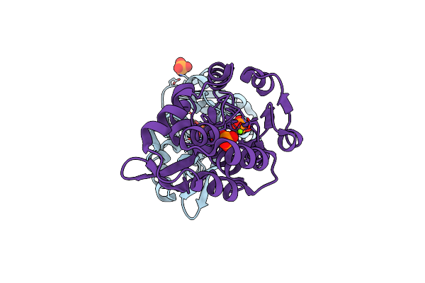 |
High Resolution Liganded Structure Of Phosphoserine Phosphatase (Pi Complex)
Organism: Methanocaldococcus jannaschii
Method: X-RAY DIFFRACTION Resolution:1.48 Å Release Date: 2002-04-03 Classification: HYDROLASE Ligands: PO4, MG |