
Search Count: 18
 |
Organism: Litorilinea aerophila
Method: ELECTRON MICROSCOPY Release Date: 2025-07-23 Classification: PROTEIN FIBRIL |
 |
Organism: Methanocaldococcus jannaschii
Method: ELECTRON MICROSCOPY Release Date: 2025-06-04 Classification: METAL BINDING PROTEIN Ligands: SF4 |
 |
Organism: Methanocaldococcus jannaschii
Method: X-RAY DIFFRACTION Release Date: 2025-06-04 Classification: METAL TRANSPORT Ligands: AN2, ZN |
 |
Organism: Methanocaldococcus jannaschii
Method: X-RAY DIFFRACTION Release Date: 2025-06-04 Classification: METAL TRANSPORT Ligands: MG, ANP |
 |
Organism: Methanocaldococcus jannaschii
Method: X-RAY DIFFRACTION Release Date: 2025-06-04 Classification: METAL TRANSPORT Ligands: EDO, SO4 |
 |
Organism: Litorilinea aerophila
Method: ELECTRON MICROSCOPY Release Date: 2025-05-21 Classification: PROTEIN FIBRIL |
 |
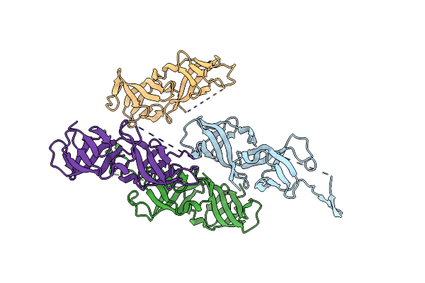
Crystal Structure Of The Hetero-Dimeric Complex From Archaeoglobus Fulgidus Prc1 And Prc2 Domains
Organism: Archaeoglobus fulgidus
Method: X-RAY DIFFRACTION Resolution:2.29 Å Release Date: 2024-04-10 Classification: CELL CYCLE |
 |
Crystal Structure Of Heterodimeric Complex Of Cdpb1 And Cdpb2 From A. Fulgidus
Organism: Archaeoglobus fulgidus
Method: X-RAY DIFFRACTION Resolution:2.29 Å Release Date: 2024-04-03 Classification: CELL CYCLE |
 |
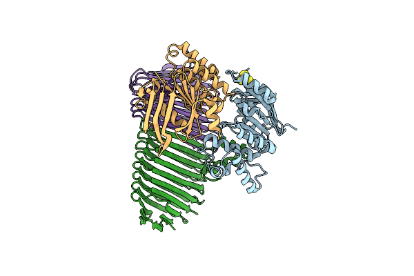
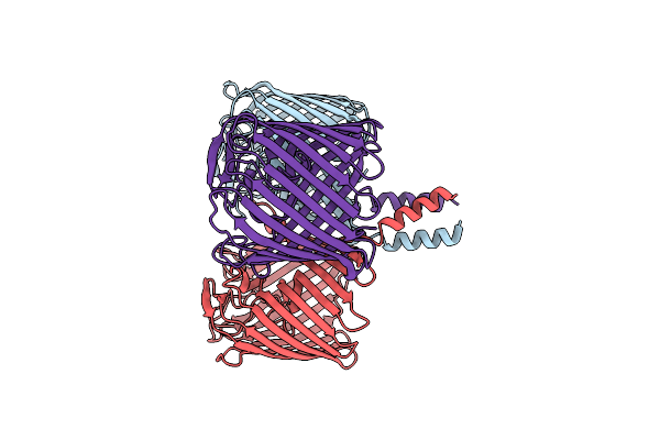
Organism: Veillonella parvula
Method: ELECTRON MICROSCOPY Release Date: 2023-11-08 Classification: MEMBRANE PROTEIN |
 |
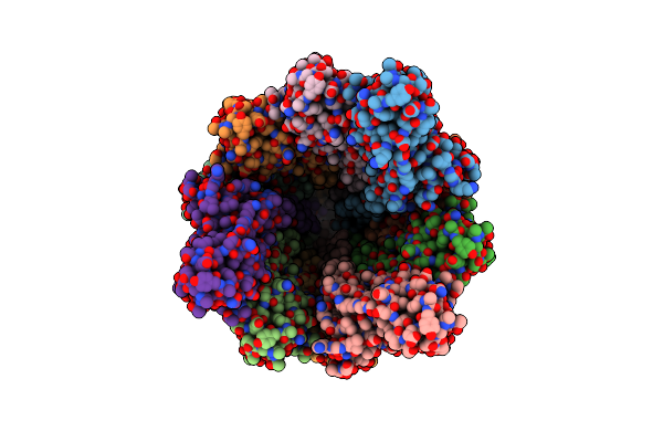
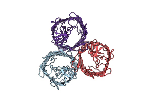
Organism: Veillonella parvula
Method: ELECTRON MICROSCOPY Release Date: 2023-11-08 Classification: MEMBRANE PROTEIN |
 |
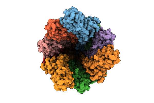
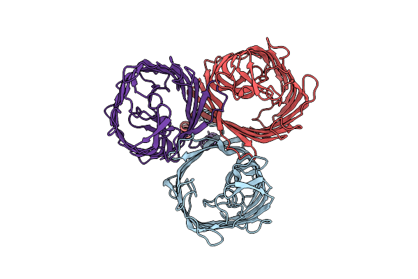
Outer Membrane Attachment Porin Ompm1 From Veillonella Parvula, C3 Symmetry
Organism: Veillonella parvula
Method: ELECTRON MICROSCOPY Release Date: 2023-11-08 Classification: MEMBRANE PROTEIN |
 |
Organism: Veillonella parvula
Method: X-RAY DIFFRACTION Resolution:1.70 Å Release Date: 2023-11-08 Classification: STRUCTURAL PROTEIN Ligands: SO4 |
 |
Organism: Methanobrevibacter smithii (strain atcc 35061 / dsm 861 / ocm 144 / ps)
Method: X-RAY DIFFRACTION Resolution:1.40 Å Release Date: 2021-03-31 Classification: CELL CYCLE Ligands: TRS |
 |
Cell Division Protein Sepf From Methanobrevibacter Smithii In Complex With Ftsz-Ctd
Organism: Methanobrevibacter smithii (strain atcc 35061 / dsm 861 / ocm 144 / ps), Methanobrevibacter smithii dsm 2375
Method: X-RAY DIFFRACTION Resolution:2.70 Å Release Date: 2021-03-31 Classification: CELL CYCLE |
 |
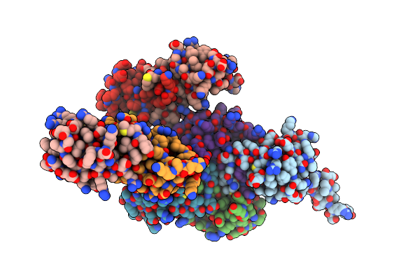
Deoxyguanylosuccinate Synthase (Dgss) Quaternary Structure With Amppcp, Dgmp, Asp, Magnesium At 2.21 Angstrom Resolution
Organism: Vibrio phage phivc8
Method: X-RAY DIFFRACTION Resolution:2.21 Å Release Date: 2020-12-16 Classification: BIOSYNTHETIC PROTEIN Ligands: SO4, DGP, ACP, ASP, MG |
 |
Deoxyguanylosuccinate Synthase (Dgss) Structure At 1.33 Angstrom Resolution.
Organism: Vibrio phage phivc8
Method: X-RAY DIFFRACTION Resolution:1.33 Å Release Date: 2019-06-12 Classification: BIOSYNTHETIC PROTEIN |
 |
Deoxyguanylosuccinate Synthase (Dgss) And Atp Structure At 1.7 Angstrom Resolution
Organism: Vibrio phage phivc8
Method: X-RAY DIFFRACTION Resolution:1.70 Å Release Date: 2019-06-12 Classification: BIOSYNTHETIC PROTEIN Ligands: ATP |
 |
Deoxyguanylosuccinate Synthase (Dgss) Quaternary Structure With Atpanddgmp At 2.3 Angstrom Resolution
Organism: Vibrio phage phivc8
Method: X-RAY DIFFRACTION Resolution:2.35 Å Release Date: 2019-06-12 Classification: BIOSYNTHETIC PROTEIN Ligands: ATP, DGP, MG, CL, GOL |