
Search Count: 31
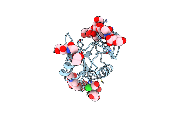 |
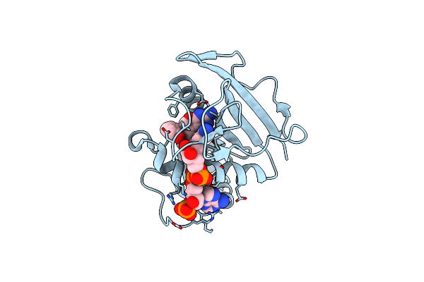
Structure Of The Murine Lyve-1 (Lymphatic Vessel Endothelial Receptor-1) Hyaluronan Binding Domain Bound With Octasaccharide Hyaluronan.
Organism: Mus musculus
Method: X-RAY DIFFRACTION Resolution:1.05 Å Release Date: 2025-02-12 Classification: CELL ADHESION Ligands: NAG, GOL, PEG, CL |
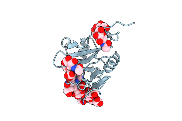 |
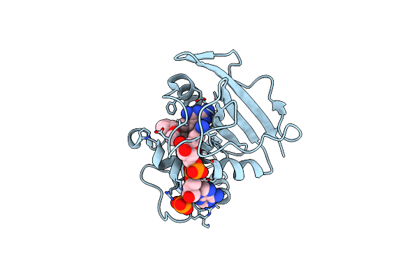
Structure Of The Human Lyve-1 (Lymphatic Vessel Endothelial Receptor-1) Hyaluronan Binding Domain Bound With Decasaccharide Hyaluronan.
Organism: Homo sapiens
Method: X-RAY DIFFRACTION Resolution:1.32 Å Release Date: 2024-11-13 Classification: CELL ADHESION Ligands: NAG |
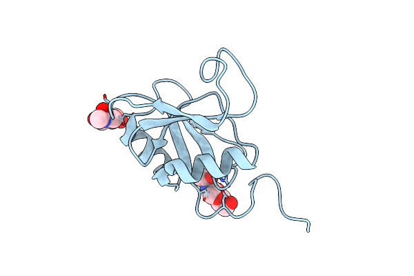 |
Structure Of The Murine Lyve-1 (Lymphatic Vessel Endothelial Receptor-1) Hyaluronan Binding Domain In An Unliganded State
Organism: Mus musculus
Method: X-RAY DIFFRACTION Resolution:1.54 Å Release Date: 2024-10-16 Classification: CELL ADHESION Ligands: NAG |
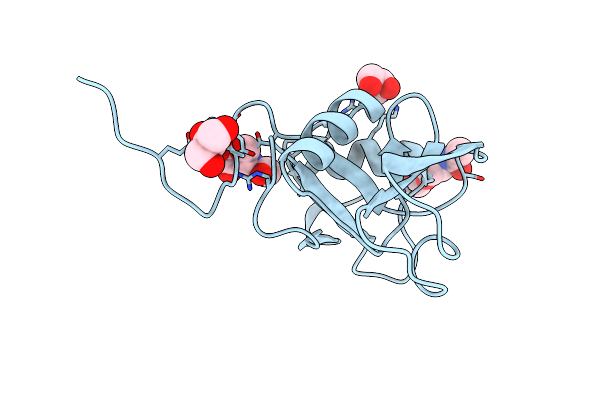 |
Structure Of The Human Lyve-1 (Lymphatic Vessel Endothelial Receptor-1) Hyaluronan Binding Domain In An Unliganded State
Organism: Homo sapiens
Method: X-RAY DIFFRACTION Resolution:1.64 Å Release Date: 2024-07-31 Classification: CELL ADHESION Ligands: NAG, GOL |
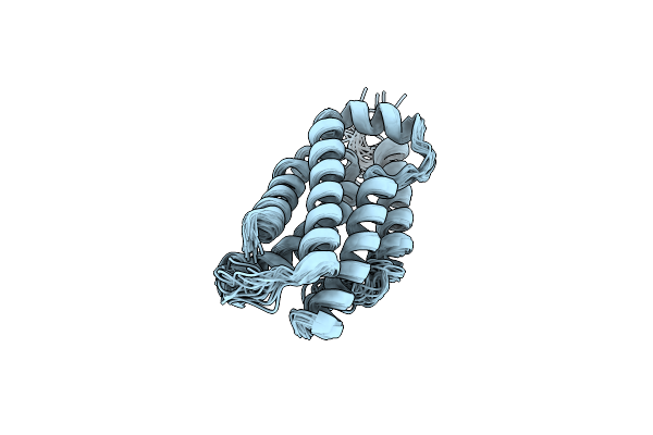 |
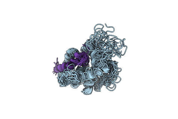
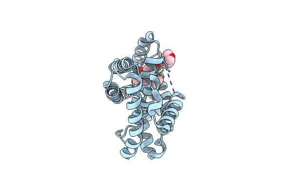
Direct Activation Of The Executioner Domain Of Mlkl By A Select Repertoire Of Inositol Phosphates
Organism: Homo sapiens
Method: SOLUTION NMR Release Date: 2019-05-15 Classification: LIPID BINDING PROTEIN |
 |
 |
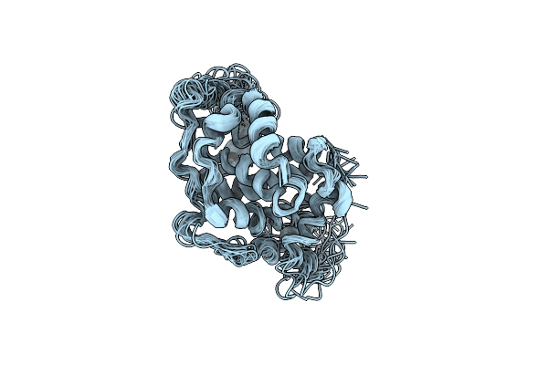
Solution Structure Of Bcl-Xl In Its P53-Bound Conformation Determined With Selective Isotope Labelling Of I,L,V Sidechains
|
 |
Solution Structure Of Bcl-Xl Containing The Alpha1-Alpha2 Disordered Loop Determined With Selective Isotope Labelling Of I,L,V Sidechains
|
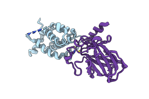 |
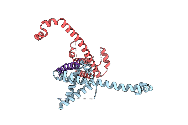
Solution Structure Of The Complex Between Bcl-Xl And The P53 Core Domain Determined With Pre Restraints
Organism: Homo sapiens
Method: SOLUTION NMR Release Date: 2014-04-30 Classification: APOPTOSIS Ligands: ZN |
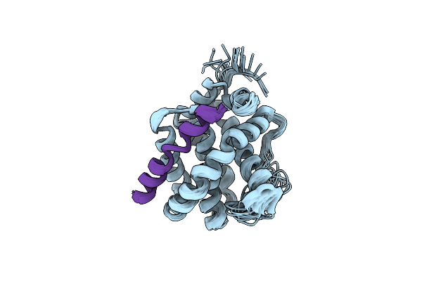 |
Organism: Homo sapiens, Synthetic construct
Method: SOLUTION NMR Release Date: 2013-04-17 Classification: APOPTOSIS |
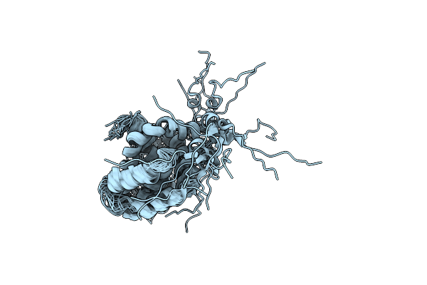 |
Solution Structure Of Bcl-Xl Determined With Selective Isotope Labelling Of I,L,V Sidechains
|
 |
Organism: Homo sapiens
Method: SOLUTION NMR Release Date: 2013-01-30 Classification: Apoptosis/PROTEIN BINDING |
 |
Crystallographic Structure Of Bcl-Xl Domain-Swapped Dimer In Complex With Puma Bh3 Peptide At 2.9A Resolution
Organism: Homo sapiens
Method: X-RAY DIFFRACTION Resolution:2.90 Å Release Date: 2013-01-23 Classification: Apoptosis/PROTEIN BINDING |
 |
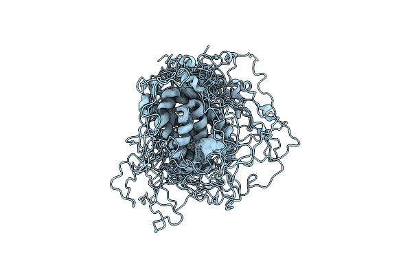
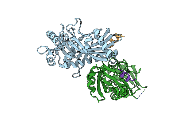
Atg8 Transfer From Atg7 To Atg3: A Distinctive E1-E2 Architecture And Mechanism In The Autophagy Pathway
Organism: Saccharomyces cerevisiae
Method: X-RAY DIFFRACTION Resolution:2.25 Å Release Date: 2011-11-23 Classification: LIGASE Ligands: ZN |
 |
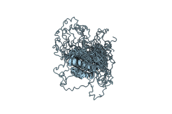
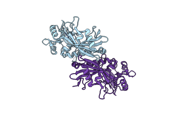
Atg8 Transfer From Atg7 To Atg3: A Distinctive E1-E2 Architecture And Mechanism In The Autophagy Pathway
Organism: Saccharomyces cerevisiae
Method: X-RAY DIFFRACTION Resolution:1.89 Å Release Date: 2011-11-23 Classification: LIGASE |
 |
Atg8 Transfer From Atg7 To Atg3: A Distinctive E1-E2 Architecture And Mechanism In The Autophagy Pathway
Organism: Saccharomyces cerevisiae
Method: X-RAY DIFFRACTION Resolution:2.08 Å Release Date: 2011-11-23 Classification: LIGASE |
 |
Atg8 Transfer From Atg7 To Atg3: A Distinctive E1-E2 Architecture And Mechanism In The Autophagy Pathway
Organism: Saccharomyces cerevisiae
Method: X-RAY DIFFRACTION Resolution:1.60 Å Release Date: 2011-11-23 Classification: LIGASE |
 |
Complex Structure Of Fxr Ligand-Binding Domain With A Tetrahydroazepinoindole Compound
Organism: Homo sapiens
Method: X-RAY DIFFRACTION Resolution:1.90 Å Release Date: 2010-03-02 Classification: TRANSCRIPTION Ligands: 635 |
 |
Alternate Binding Modes Observed For The E- And Z-Isomers Of 2,4-Diaminofuro[2,3D]Pyrimidines As Ternary Complexes With Nadph And Mouse Dihydrofolate Reductase
Organism: Mus musculus
Method: X-RAY DIFFRACTION Resolution:1.60 Å Release Date: 2009-10-13 Classification: OXIDOREDUCTASE Ligands: NDP, 51P |
 |
Alternate Binding Modes Observed For The E- And Z-Isomers Of 2,4-Diaminofuro[2,3-D]Pyrimidines As Ternary Complexes With Nadph And Mouse Dihydrofolate Reductase
Organism: Mus musculus
Method: X-RAY DIFFRACTION Resolution:2.05 Å Release Date: 2009-10-13 Classification: OXIDOREDUCTASE Ligands: D09, NDP |