
Search Count: 24
 |
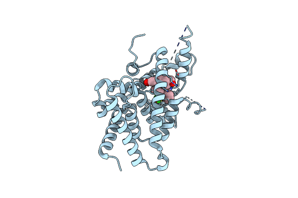
Organism: Homo sapiens
Method: X-RAY DIFFRACTION Resolution:2.37 Å Release Date: 2021-11-03 Classification: ONCOPROTEIN Ligands: 5I4 |
 |
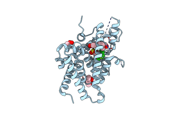
Organism: Homo sapiens
Method: X-RAY DIFFRACTION Resolution:2.15 Å Release Date: 2021-01-13 Classification: NUCLEAR PROTEIN Ligands: EDO |
 |
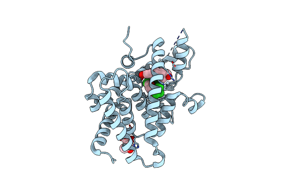
Organism: Homo sapiens
Method: X-RAY DIFFRACTION Resolution:2.25 Å Release Date: 2021-01-13 Classification: NUCLEAR PROTEIN Ligands: S6H, IPA, GOL |
 |
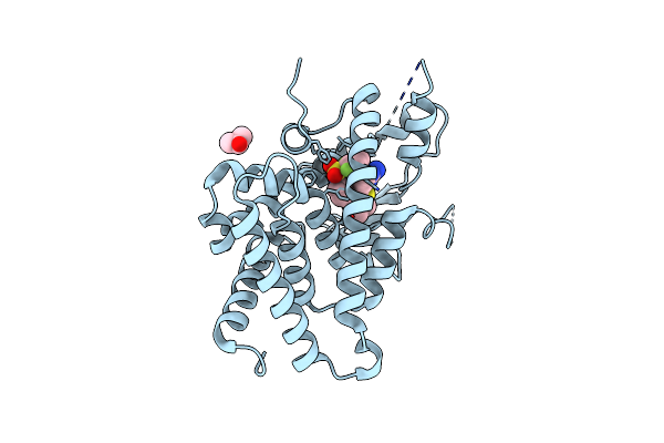
Organism: Homo sapiens
Method: X-RAY DIFFRACTION Resolution:2.26 Å Release Date: 2021-01-13 Classification: NUCLEAR PROTEIN Ligands: CL6 |
 |
Organism: Homo sapiens
Method: X-RAY DIFFRACTION Resolution:2.55 Å Release Date: 2021-01-13 Classification: NUCLEAR PROTEIN Ligands: MPD, S68, GOL |
 |
Organism: Homo sapiens
Method: X-RAY DIFFRACTION Resolution:2.05 Å Release Date: 2021-01-13 Classification: NUCLEAR PROTEIN Ligands: 27H, GOL, IPA |
 |
Organism: Homo sapiens
Method: X-RAY DIFFRACTION Resolution:2.65 Å Release Date: 2021-01-13 Classification: NUCLEAR PROTEIN Ligands: S6W, IPA |
 |
Organism: Homo sapiens
Method: X-RAY DIFFRACTION Resolution:1.90 Å Release Date: 2021-01-13 Classification: NUCLEAR PROTEIN Ligands: GOL, S6Z, IPA |
 |
Organism: Homo sapiens
Method: X-RAY DIFFRACTION Resolution:2.45 Å Release Date: 2021-01-13 Classification: NUCLEAR PROTEIN Ligands: S6T, IPA |
 |
Organism: Homo sapiens
Method: X-RAY DIFFRACTION Resolution:2.70 Å Release Date: 2021-01-13 Classification: NUCLEAR PROTEIN Ligands: TBY |
 |
Organism: Homo sapiens
Method: X-RAY DIFFRACTION Resolution:2.55 Å Release Date: 2021-01-13 Classification: NUCLEAR PROTEIN Ligands: IPA, MPD, 27J |
 |
Crystal Structure Of The Hpxr-Lbd In Complex With Estradiol And Cis-Chlordane
Organism: Homo sapiens
Method: X-RAY DIFFRACTION Resolution:2.15 Å Release Date: 2021-01-13 Classification: NUCLEAR PROTEIN Ligands: EST, MPD, S6E, S6H, IPA |
 |
Crystal Structure Of The Hpxr-Lbd In Complex With Estradiol And Clotrimazole
Organism: Homo sapiens
Method: X-RAY DIFFRACTION Resolution:2.30 Å Release Date: 2021-01-13 Classification: NUCLEAR PROTEIN Ligands: CL6, EST |
 |
Organism: Homo sapiens
Method: X-RAY DIFFRACTION Resolution:2.00 Å Release Date: 2021-01-13 Classification: NUCLEAR PROTEIN Ligands: EST, MPD, S68, GOL |
 |
Crystal Structure Of The Hpxr-Lbd In Complex With Estradiol And Heptachlor Endo-Epoxide
Organism: Homo sapiens
Method: X-RAY DIFFRACTION Resolution:2.50 Å Release Date: 2021-01-13 Classification: NUCLEAR PROTEIN Ligands: EST, S65, MPD |
 |
Organism: Homo sapiens
Method: X-RAY DIFFRACTION Resolution:2.28 Å Release Date: 2020-03-25 Classification: NUCLEAR PROTEIN Ligands: P06, IPA |
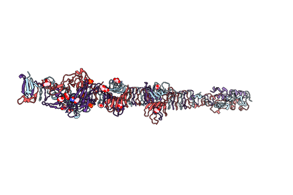 |
Crystal Structure Of The Carboxy-Terminal Region Of The Bacteriophage T4 Proximal Long Tail Fibre Protein Gp34, Residues 795 To 1289, At 1.9 Angstrom.
Organism: Enterobacteria phage t4
Method: X-RAY DIFFRACTION Resolution:1.90 Å Release Date: 2017-07-12 Classification: VIRAL PROTEIN Ligands: GOL, PO4, ACT, NA, URE |
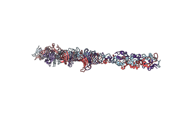 |
Crystal Structure Of The Carboxy-Terminal Region Of The Bacteriophage T4 Proximal Long Tail Fibre Protein Gp34, Residues 744-1289 At 2.9 Angstrom Resolution
Organism: Enterobacteria phage t4
Method: X-RAY DIFFRACTION Resolution:2.89 Å Release Date: 2017-07-12 Classification: VIRAL PROTEIN Ligands: GOL |
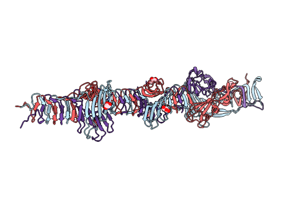 |
Crystal Structure Of The Carboxy-Terminal Region Of The Bacteriophage T4 Proximal Long Tail Fibre Protein Gp34, P21 Native Crystal
Organism: Enterobacteria phage t4
Method: X-RAY DIFFRACTION Resolution:2.00 Å Release Date: 2015-12-16 Classification: VIRAL PROTEIN Ligands: GOL |
 |
Structure Of The Carboxy-Terminal Domain Of The Bacteriophage T5 L- Shaped Tail Fibre
Organism: Escherichia phage t5
Method: X-RAY DIFFRACTION Resolution:2.30 Å Release Date: 2015-12-16 Classification: VIRAL PROTEIN |