
Search Count: 20
 |
Organism: Hypocrea jecorina
Method: X-RAY DIFFRACTION, NEUTRON DIFFRACTION Release Date: 2015-09-30 Classification: HYDROLASE Ligands: IOD, DOD |
 |
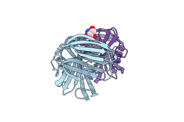
Joint X-Ray/Neutron Structure Of Trichoderma Reesei Xylanase Ii In Complex With Mes At Ph 5.7
Organism: Trichoderma reesei
Method: X-RAY DIFFRACTION, NEUTRON DIFFRACTION Release Date: 2015-09-23 Classification: HYDROLASE Ligands: IOD, MES, DOD |
 |
Organism: Trichoderma reesei
Method: X-RAY DIFFRACTION, NEUTRON DIFFRACTION Resolution:1.70 Å, 2.00 Å Release Date: 2015-09-23 Classification: HYDROLASE Ligands: IOD, DOD |
 |
Organism: Trichoderma reesei
Method: X-RAY DIFFRACTION, NEUTRON DIFFRACTION Resolution:1.60 Å, 2.00 Å Release Date: 2015-09-23 Classification: HYDROLASE Ligands: IOD, DOD |
 |
Organism: Trichoderma reesei
Method: X-RAY DIFFRACTION, NEUTRON DIFFRACTION Resolution:1.60 Å, 1.70 Å Release Date: 2015-09-23 Classification: HYDROLASE Ligands: IOD, DOD |
 |
Organism: Hypocrea jecorina
Method: X-RAY DIFFRACTION Resolution:1.25 Å Release Date: 2015-09-23 Classification: HYDROLASE Ligands: IOD |
 |
Organism: Hypocrea jecorina
Method: X-RAY DIFFRACTION Resolution:1.50 Å Release Date: 2015-09-23 Classification: HYDROLASE Ligands: IOD, TRS |
 |
Organism: Hypocrea jecorina
Method: X-RAY DIFFRACTION Resolution:1.50 Å Release Date: 2015-09-23 Classification: HYDROLASE Ligands: IOD, MES |
 |
Joint X-Ray And Neutron Structure Of Streptomyces Rubiginosus D-Xylose Isomerase In Complex With Two Cd2+ Ions And Cyclic Beta-L-Arabinose
Organism: Streptomyces rubiginosus
Method: NEUTRON DIFFRACTION, X-RAY DIFFRACTION Resolution:2.00 Å Release Date: 2014-09-03 Classification: ISOMERASE Ligands: CD, ARB, DOD |
 |
Joint X-Ray And Neutron Structure Of Streptomyces Rubiginosus D-Xylose Isomerase In Complex With Two Ni2+ Ions And Linear L-Arabinose
Organism: Streptomyces rubiginosus
Method: X-RAY DIFFRACTION, NEUTRON DIFFRACTION Resolution:1.80 Å Release Date: 2014-09-03 Classification: ISOMERASE Ligands: NI, LAI, DOD |
 |
Room Temperature X-Ray Structure Of D-Xylose Isomerase In Complex With Two Cd2+ Ions And L-Ribulose
Organism: Streptomyces rubiginosus
Method: X-RAY DIFFRACTION Resolution:1.55 Å Release Date: 2014-09-03 Classification: ISOMERASE Ligands: CD, RUU |
 |
Room Temperature X-Ray Structure Of D-Xylose Isomerase In Complex With Two Ni2+ Ions And L-Ribulose
Organism: Streptomyces rubiginosus
Method: X-RAY DIFFRACTION Resolution:1.70 Å Release Date: 2014-09-03 Classification: ISOMERASE Ligands: NI, 34V |
 |
Room Temperature X-Ray Structure Of D-Xylose Isomerase In Complex With Two Mg2+ Ions And L-Ribulose
Organism: Streptomyces rubiginosus
Method: X-RAY DIFFRACTION Resolution:1.56 Å Release Date: 2014-09-03 Classification: ISOMERASE Ligands: MG, RUU |
 |
Room Temperature X-Ray Structure Of D-Xylose Isomerase In Complex With Two Ni2+ Ions And L-Ribose
Organism: Streptomyces rubiginosus
Method: X-RAY DIFFRACTION Resolution:1.60 Å Release Date: 2014-09-03 Classification: ISOMERASE Ligands: NI, Z6J |
 |
Room Temperature X-Ray Structure Of D-Xylose Isomerase In Complex With Two Mg2+ Ions And L-Ribose
Organism: Streptomyces rubiginosus
Method: X-RAY DIFFRACTION Resolution:1.55 Å Release Date: 2014-09-03 Classification: ISOMERASE Ligands: MG, 32O |
 |
Crystal Structure Of 3-Isopropylmalate Dehydratase (Leud)From Methhanocaldococcus Jannaschii Dsm2661 (Mj1271)
Organism: Methanocaldococcus jannaschii
Method: X-RAY DIFFRACTION Resolution:2.10 Å Release Date: 2008-04-22 Classification: LYASE Ligands: ZN, PEG |
 |
Organism: Methanocaldococcus jannaschii
Method: X-RAY DIFFRACTION Resolution:1.60 Å Release Date: 2003-12-09 Classification: LYASE Ligands: SO4 |
 |
The Crystal Structure Of Pyruvoyl-Dependent Arginine Decarboxylase From Methanococcus Jannaschii
Organism: Methanocaldococcus jannaschii
Method: X-RAY DIFFRACTION Resolution:2.20 Å Release Date: 2003-03-25 Classification: LYASE Ligands: AG2 |
 |
The Crystal Structure Of Pyruvoyl-Dependent Arginine Decarboxylase From Methanococcus Jannashii
Organism: Methanocaldococcus jannaschii
Method: X-RAY DIFFRACTION Resolution:1.40 Å Release Date: 2003-03-25 Classification: LYASE Ligands: AG2, MRD |
 |
Organism: Methanocaldococcus jannaschii
Method: X-RAY DIFFRACTION Resolution:1.90 Å Release Date: 2003-03-25 Classification: LYASE Ligands: MRD |