
Search Count: 35
 |
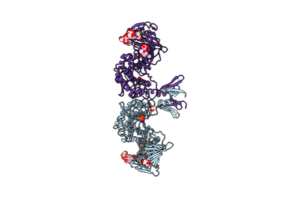
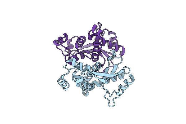
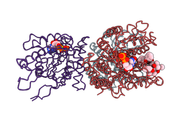
Topological Model Of The P2 Virion Baseplate In Activated Conformation (Closed Tal Trimer)
Organism: Lactococcus phage p2
Method: ELECTRON MICROSCOPY Release Date: 2020-08-26 Classification: VIRAL PROTEIN |
 |
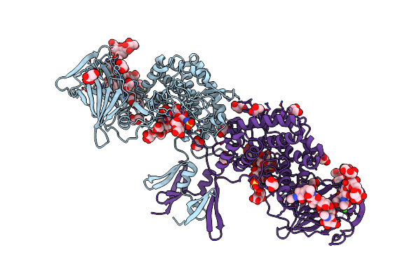
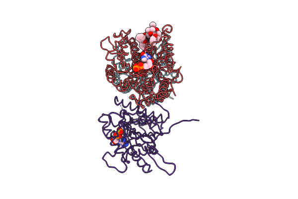
Organism: Lactococcus phage p2
Method: ELECTRON MICROSCOPY Release Date: 2020-08-26 Classification: VIRAL PROTEIN |
 |
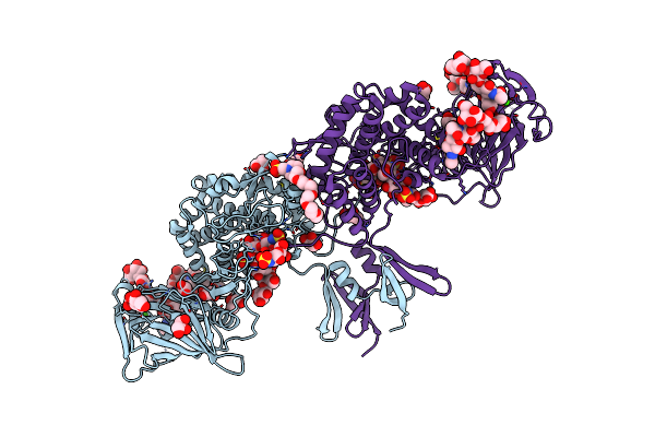
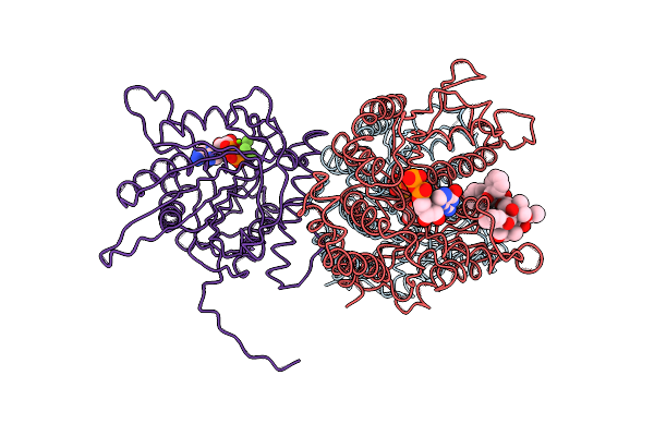
Organism: Lactococcus phage p2
Method: ELECTRON MICROSCOPY Release Date: 2020-08-26 Classification: VIRAL PROTEIN |
 |
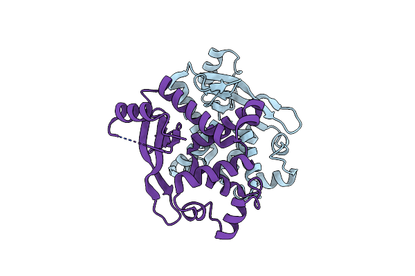
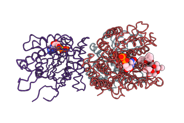
Organism: Streptococcus phage d1811, Streptococcus thermophilus (strain atcc baa-491 / lmd-9), Streptococcus thermophilus, Streptococcus virus 2972
Method: ELECTRON MICROSCOPY Resolution:3.20 Å Release Date: 2019-10-02 Classification: HYDROLASE |
 |
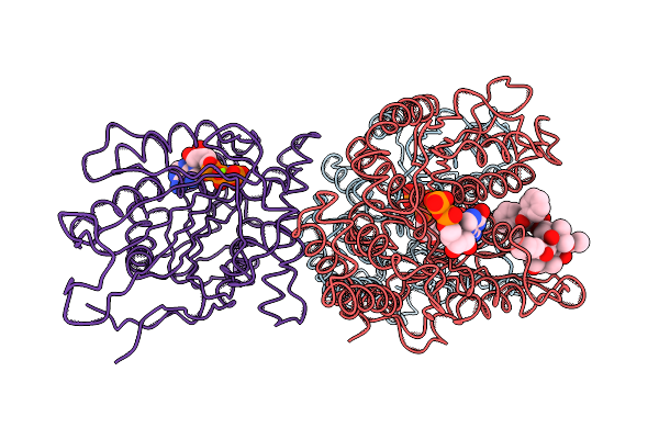
Organism: Streptococcus phage d1811, Streptococcus thermophilus (strain atcc baa-491 / lmd-9), Streptococcus thermophilus, Streptococcus virus 2972
Method: ELECTRON MICROSCOPY Resolution:3.00 Å Release Date: 2019-10-02 Classification: HYDROLASE |
 |
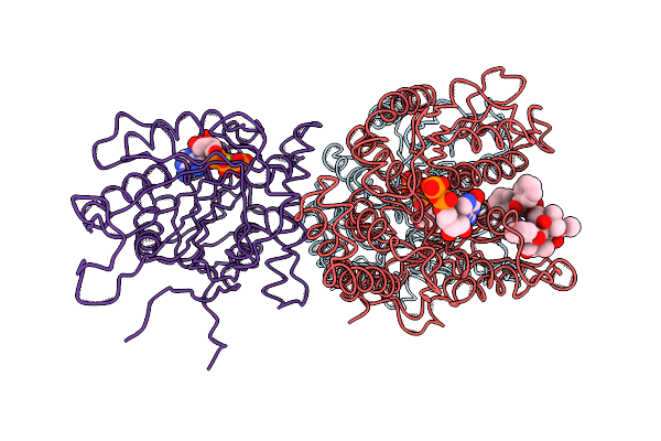
Organism: Streptococcus thermophilus dgcc 7710, Streptococcus phage d1811, Streptococcus phage 2972
Method: ELECTRON MICROSCOPY Release Date: 2019-10-02 Classification: HYDROLASE |
 |
Organism: Streptococcus phage d1811, Streptococcus thermophilus dgcc 7710, Streptococcus thermophilus, Streptococcus virus 2972
Method: ELECTRON MICROSCOPY Release Date: 2019-10-02 Classification: HYDROLASE |
 |
Organism: Homo sapiens
Method: X-RAY DIFFRACTION Resolution:2.52 Å Release Date: 2019-04-03 Classification: ISOMERASE Ligands: NAG, ACT, SO4, CA, CL |
 |
Organism: Homo sapiens
Method: X-RAY DIFFRACTION Resolution:2.10 Å Release Date: 2019-04-03 Classification: ISOMERASE Ligands: NAG, GOL, ACT, CA |
 |
Organism: Homo sapiens
Method: X-RAY DIFFRACTION Resolution:2.45 Å Release Date: 2019-04-03 Classification: ISOMERASE Ligands: CA, NAG, GOL, MES, P6G |
 |
Organism: Streptococcus phage dt1
Method: X-RAY DIFFRACTION Resolution:1.96 Å Release Date: 2018-06-06 Classification: VIRAL PROTEIN |
 |
Organism: Streptococcus phage 73
Method: X-RAY DIFFRACTION Resolution:2.50 Å Release Date: 2018-06-06 Classification: VIRAL PROTEIN |
 |
Pseudo-Atomic Model Of Microtubule-Bound S.Pombe Kinesin-5 Motor Domain In The Amppnp State (Based On Cryo-Electron Microscopy Experiment): The N-Terminus Conformation Allows Formation Of A Cover Neck Bundle.
Organism: Schizosaccharomyces pombe (strain 972 / atcc 24843), Bos taurus
Method: ELECTRON MICROSCOPY Release Date: 2016-11-30 Classification: MOTOR PROTEIN Ligands: MG, GTP, GDP, TA1, ANP |
 |
Pseudo-Atomic Model Of Microtubule-Bound S. Pombe Kinesin-5 Motor Domain In The Amppnp State (Based On Cryo-Electron Microscopy Experiment): The N-Terminus Adopts Multiple Conformations
Organism: Schizosaccharomyces pombe (strain 972 / atcc 24843), Bos taurus
Method: ELECTRON MICROSCOPY Release Date: 2016-11-30 Classification: MOTOR PROTEIN Ligands: MG, GTP, GDP, TA1, ANP |
 |
Pseudo-Atomic Model Of Microtubule-Bound S.Pombe Kinesin-5 Motor Domain In The Amppnp State (Based On Cryo-Electron Microscopy Experiment): The N-Terminus Adopts Multiple Conformations.
Organism: Schizosaccharomyces pombe (strain 972 / atcc 24843), Bos taurus
Method: ELECTRON MICROSCOPY Release Date: 2016-11-30 Classification: MOTOR PROTEIN Ligands: MG, GTP, GDP, TA1, ANP |
 |
Organism: Toscana virus
Method: X-RAY DIFFRACTION Resolution:3.70 Å Release Date: 2016-03-09 Classification: VIRAL PROTEIN |
 |
Pseudo-Atomic Model Of Microtubule-Bound Human Kinesin-5 Motor Domain In The Adp State, Based On Cryo-Electron Microscopy Experiment.
Organism: Homo sapiens, Bos taurus
Method: ELECTRON MICROSCOPY Resolution:10.00 Å Release Date: 2014-02-05 Classification: MOTOR PROTEIN Ligands: GTP, GDP, TA1, MG, ADP |
 |
Pseudo-Atomic Model Of Microtubule-Bound Human Kinesin-5 Motor Domain In The Adp.Alfx State, Based On Cryo-Electron Microscopy Experiment.
Organism: Homo sapiens, Bos taurus
Method: ELECTRON MICROSCOPY Resolution:9.20 Å Release Date: 2014-02-05 Classification: MOTOR PROTEIN Ligands: MG, GTP, GDP, TA1, AF3, ADP |