
Search Count: 49
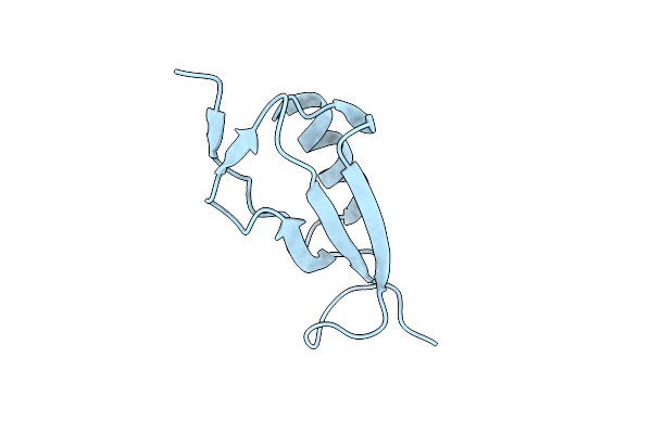 |
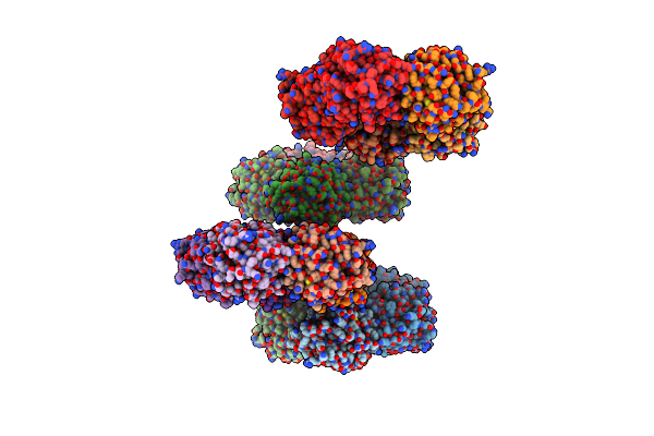
Pasta Subunit 4 Of Streptococcus Pneumoniae Stkp Crystallized With Peg And Succinate
Organism: Streptococcus pneumoniae (strain atcc baa-255 / r6)
Method: X-RAY DIFFRACTION Resolution:1.90 Å Release Date: 2017-11-08 Classification: TRANSFERASE |
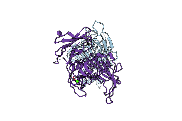 |
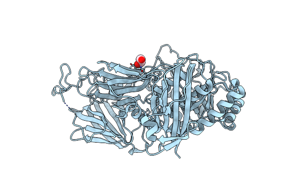
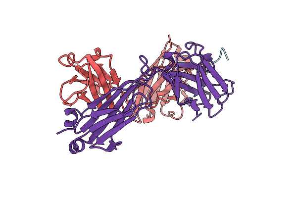
Crystal Structure Of Pectate Lyase Pel3 From Pectobacterium Carotovorum With Two Monomers In The A.U
Organism: Pectobacterium carotovorum
Method: X-RAY DIFFRACTION Resolution:1.80 Å Release Date: 2015-08-05 Classification: LYASE Ligands: CA |
 |
Crystal Structure Of Pectate Lyase Pel3 From Pectobacterium Carotovorum With One Monomer In The A.U.
Organism: Pectobacterium carotovorum
Method: X-RAY DIFFRACTION Resolution:2.10 Å Release Date: 2015-08-05 Classification: LYASE Ligands: SO4, EDO |
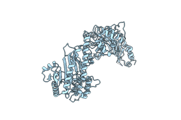 |
Organism: Schistosoma mansoni
Method: X-RAY DIFFRACTION Resolution:2.20 Å Release Date: 2015-04-15 Classification: TRANSFERASE |
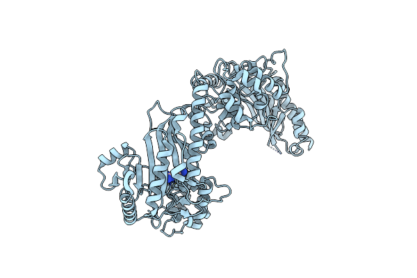 |
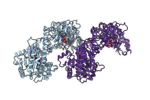
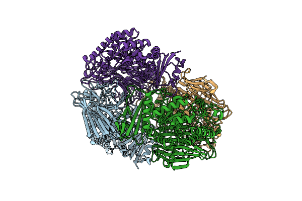
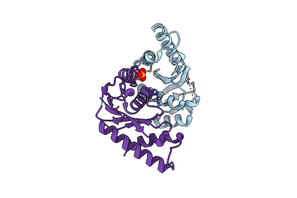
The Duplicated Taurocyamine Kinase From Schistosoma Mansoni Complexed With Arginine
Organism: Schistosoma mansoni
Method: X-RAY DIFFRACTION Resolution:1.90 Å Release Date: 2015-04-15 Classification: TRANSFERASE Ligands: ARG |
 |
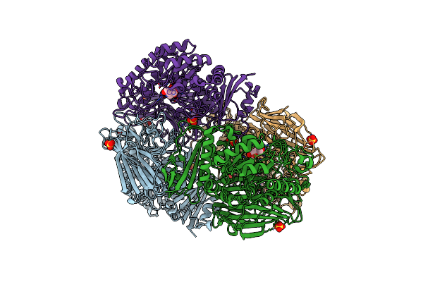
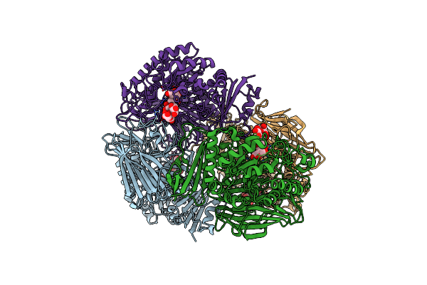
The Duplicated Taurocyamine Kinase From Schistosoma Mansoni With Bound Transition State Analog (Tsa) Components
Organism: Schistosoma mansoni
Method: X-RAY DIFFRACTION Resolution:2.30 Å Release Date: 2015-04-15 Classification: TRANSFERASE Ligands: ADP, 3S5, MG, NO3 |
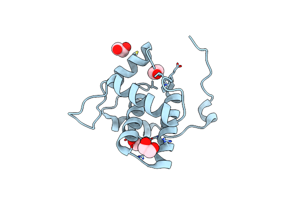 |
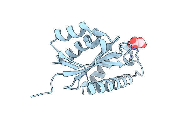
Crystal Structure Of The Full-Length Matrix Subunit (P15) Of The Feline Immunodeficiency Virus (Fiv) Gag Polyprotein
Organism: Feline immunodeficiency virus
Method: X-RAY DIFFRACTION Resolution:2.00 Å Release Date: 2013-07-10 Classification: VIRAL PROTEIN Ligands: EDO |
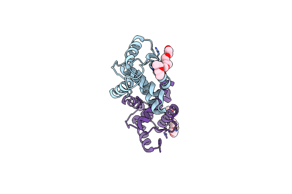 |
Crystal Structure Of A C-Terminal Truncated Form Of The Matrix Subunit (P15) Of Feline Immunodeficiency Virus (Fiv)
Organism: Feline immunodeficiency virus
Method: X-RAY DIFFRACTION Resolution:2.70 Å Release Date: 2013-07-10 Classification: VIRAL PROTEIN Ligands: 7PE |
 |
Crystal Structure Of Gh36 Alpha-Galactosidase Agaa A355E From Geobacillus Stearothermophilus
Organism: Geobacillus stearothermophilus
Method: X-RAY DIFFRACTION Resolution:2.80 Å Release Date: 2012-10-03 Classification: HYDROLASE Ligands: SO4 |
 |
Crystal Structure Of Gh36 Alpha-Galactosidase Agab From Geobacillus Stearothermophilus
Organism: Geobacillus stearothermophilus
Method: X-RAY DIFFRACTION Resolution:1.80 Å Release Date: 2012-10-03 Classification: HYDROLASE Ligands: EDO |
 |
Crystal Structure Of Gh36 Alpha-Galactosidase Agaa From Geobacillus Stearothermophilus
Organism: Geobacillus stearothermophilus
Method: X-RAY DIFFRACTION Resolution:3.20 Å Release Date: 2012-10-03 Classification: HYDROLASE |
 |
Crystal Structure Of Gh36 Alpha-Galactosidase Agaa A355E From Geobacillus Stearothermophilus In Complex With 1-Deoxygalactonojirimycin
Organism: Geobacillus stearothermophilus
Method: X-RAY DIFFRACTION Resolution:2.60 Å Release Date: 2012-10-03 Classification: HYDROLASE Ligands: DGJ, SO4, EDO |
 |
Crystal Structure Of Gh36 Alpha-Galactosidase Agaa A355E D548N From Geobacillus Stearothermophilus In Complex With Raffinose
Organism: Geobacillus stearothermophilus
Method: X-RAY DIFFRACTION Resolution:2.60 Å Release Date: 2012-10-03 Classification: HYDROLASE |
 |
Crystal Structure Of Gh36 Alpha-Galactosidase Agaa A355E D478A From Geobacillus Stearothermophilus In Complex With Stachyose
Organism: Geobacillus stearothermophilus
Method: X-RAY DIFFRACTION Resolution:3.60 Å Release Date: 2012-10-03 Classification: HYDROLASE |
 |
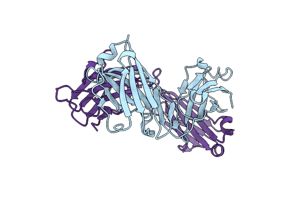
Organism: Homo sapiens
Method: X-RAY DIFFRACTION Resolution:1.98 Å Release Date: 2012-01-11 Classification: IMMUNE SYSTEM Ligands: CA, ZN |
 |
Organism: Homo sapiens
Method: X-RAY DIFFRACTION Resolution:3.00 Å Release Date: 2012-01-11 Classification: IMMUNE SYSTEM Ligands: CA |
 |
Crystal Structure Of The Rous Associated Virus Integrase Catalytic Domain In Mes Buffer Ph 6.0
Organism: Rous sarcoma virus
Method: X-RAY DIFFRACTION Resolution:1.80 Å Release Date: 2011-08-31 Classification: DNA BINDING PROTEIN Ligands: MES, ZN |
 |
Crystal Structure Of The Rous Associated Virus Integrase Catalytic Domain A182T In Citrate Buffer Ph 6.2
Organism: Rous sarcoma virus
Method: X-RAY DIFFRACTION Resolution:1.55 Å Release Date: 2011-08-31 Classification: DNA BINDING PROTEIN Ligands: FLC |
 |
Organism: Mus musculus
Method: X-RAY DIFFRACTION Resolution:2.00 Å Release Date: 2010-11-10 Classification: IMMUNE SYSTEM |
 |
Organism: Mus musculus
Method: X-RAY DIFFRACTION Resolution:2.10 Å Release Date: 2010-11-10 Classification: IMMUNE SYSTEM |