
Search Count: 37
 |
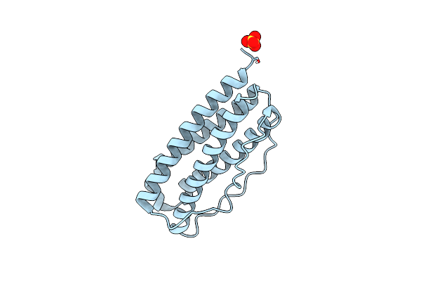
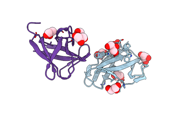
The Crystal Structure Of Human Apolipoprotein A-I In Complex With Fab 55201
Organism: Homo sapiens, Mus musculus
Method: X-RAY DIFFRACTION Release Date: 2025-08-27 Classification: LIPID TRANSPORT/IMMUNE SYSTEM |
 |
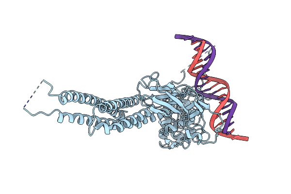
Crystal Structure Of Lyssavirus Rabies (Ni-Ce Strain) Nucleoprotein In Complex With Phosphoprotein Chaperone
Organism: Rabies virus (strain nishigahara rceh)
Method: X-RAY DIFFRACTION Release Date: 2025-04-23 Classification: VIRAL PROTEIN |
 |
Crystal Structure Of Lyssavirus Rabies (Nishigahara Strain) Nucleoprotein In Complex With Phosphomimetic Phosphoprotein S48E
Organism: Lyssavirus rabies
Method: X-RAY DIFFRACTION Release Date: 2025-04-23 Classification: VIRAL PROTEIN |
 |
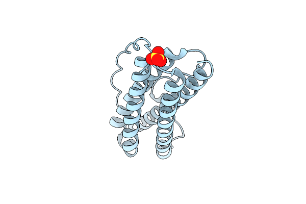
Organism: Homo sapiens
Method: ELECTRON MICROSCOPY Release Date: 2025-03-26 Classification: IMMUNE SYSTEM |
 |
Organism: Homo sapiens
Method: ELECTRON MICROSCOPY Resolution:3.07 Å Release Date: 2025-03-26 Classification: IMMUNE SYSTEM |
 |
Organism: Homo sapiens
Method: ELECTRON MICROSCOPY Release Date: 2023-11-29 Classification: CYTOKINE Ligands: NAG |
 |
The Structure Of The Il-11 Signalling Complex, With Full-Length Extracellular Gp130
Organism: Homo sapiens
Method: ELECTRON MICROSCOPY Release Date: 2023-11-29 Classification: CYTOKINE Ligands: NAG |
 |
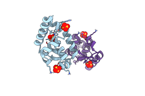
Organism: Homo sapiens
Method: X-RAY DIFFRACTION Resolution:3.78 Å Release Date: 2023-11-29 Classification: CYTOKINE Ligands: NAG |
 |
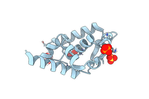
Organism: Homo sapiens
Method: X-RAY DIFFRACTION Resolution:1.48 Å Release Date: 2023-11-29 Classification: CYTOKINE Ligands: SO4, CL |
 |
Organism: Homo sapiens
Method: X-RAY DIFFRACTION Resolution:1.80 Å Release Date: 2023-11-29 Classification: CYTOKINE Ligands: SO4 |
 |
Organism: Rabies virus nishigahara rceh
Method: X-RAY DIFFRACTION Resolution:1.70 Å Release Date: 2022-04-20 Classification: VIRAL PROTEIN Ligands: SO4 |
 |
Organism: Rabies virus nishigahara rceh
Method: X-RAY DIFFRACTION Resolution:1.50 Å Release Date: 2022-04-20 Classification: VIRAL PROTEIN Ligands: EDO, SO4, PO4 |
 |
Crystal Structure Of Rabies Virus (Nishigahara Strain) Phosphoprotein C-Terminal Domain (K214A)
Organism: Rabies virus (strain nishigahara rceh)
Method: X-RAY DIFFRACTION Resolution:3.00 Å Release Date: 2021-03-17 Classification: VIRAL PROTEIN |
 |
Organism: Duvenhage virus
Method: X-RAY DIFFRACTION Resolution:1.95 Å Release Date: 2021-03-17 Classification: VIRAL PROTEIN |
 |
Organism: Homo sapiens
Method: X-RAY DIFFRACTION Release Date: 2020-08-19 Classification: TRANSCRIPTION |
 |
Organism: Homo sapiens
Method: X-RAY DIFFRACTION Resolution:1.62 Å Release Date: 2020-05-06 Classification: PROTEIN BINDING Ligands: SO4, CL |
 |
Organism: Homo sapiens
Method: X-RAY DIFFRACTION Resolution:3.43 Å Release Date: 2020-05-06 Classification: PROTEIN BINDING Ligands: NAG, SO4 |
 |
Beta1 Carbohydrate Binding Module (Cbm) Of Amp-Activated Protein Kinase (Ampk) In Complex With Glucosyl-Beta-Cyclododextrin
Organism: Rattus norvegicus
Method: X-RAY DIFFRACTION Resolution:1.72 Å Release Date: 2015-06-24 Classification: SUGAR BINDING PROTEIN Ligands: GOL, SO4 |
 |
Structure Of Amylase Binding Protein A Of Streptococcous Gordonii: A Potential Receptor For Human Salivary Amylase Enzyme
Organism: Streptococcus gordonii str. challis
Method: SOLUTION NMR Release Date: 2015-05-13 Classification: HYDROLASE RECEPTOR |
 |
Beta2 Carbohydrate Binding Module (Cbm) Of Amp-Activated Protein Kinase (Ampk)
Organism: Rattus norvegicus
Method: X-RAY DIFFRACTION Resolution:1.60 Å Release Date: 2015-04-08 Classification: PROTEIN BINDING Ligands: GOL |