
Search Count: 22
 |
Organism: Homo sapiens
Method: ELECTRON MICROSCOPY Release Date: 2025-02-19 Classification: MEMBRANE PROTEIN Ligands: CLR, PCW, LPE, P5S |
 |
Organism: Homo sapiens
Method: ELECTRON MICROSCOPY Release Date: 2025-02-19 Classification: MEMBRANE PROTEIN Ligands: CLR, PCW, LPE, P5S |
 |
Organism: Homo sapiens
Method: ELECTRON MICROSCOPY Release Date: 2025-02-19 Classification: MEMBRANE PROTEIN Ligands: CLR, PCW, LPE, P5S |
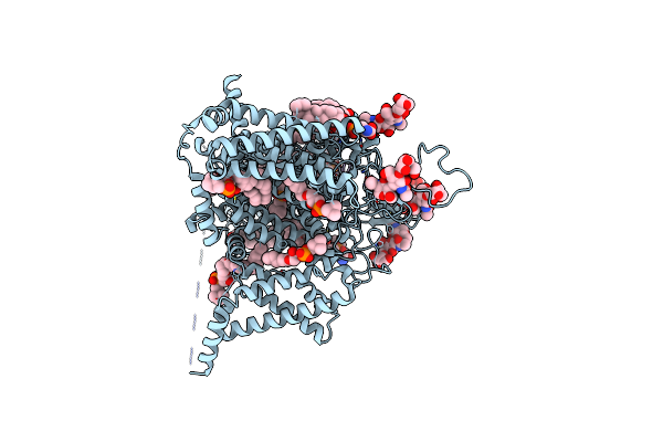 |
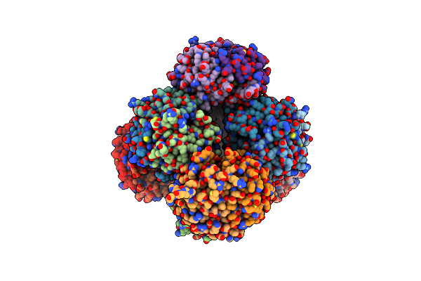
Tarantula Venom Peptide Protoxin-I Bound To Full-Length Human Voltage-Gated Sodium Channel 1.8 (Nav1.8)
Organism: Homo sapiens, Thrixopelma pruriens
Method: ELECTRON MICROSCOPY Release Date: 2025-02-19 Classification: MEMBRANE PROTEIN Ligands: CLR, PCW, LPE, P5S |
 |
Organism: Synthetic construct
Method: ELECTRON MICROSCOPY Release Date: 2024-06-05 Classification: DE NOVO PROTEIN |
 |
Organism: Synthetic construct
Method: ELECTRON MICROSCOPY Release Date: 2024-06-05 Classification: DE NOVO PROTEIN |
 |
Organism: Thermotoga maritima msb8
Method: ELECTRON MICROSCOPY Release Date: 2023-01-11 Classification: DE NOVO PROTEIN |
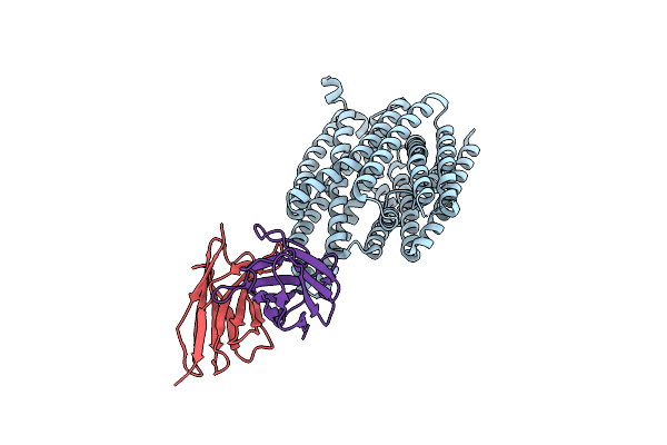 |
Organism: Homo sapiens, Mus musculus
Method: ELECTRON MICROSCOPY Release Date: 2020-09-09 Classification: TRANSPORT PROTEIN/IMMUNE SYSTEM |
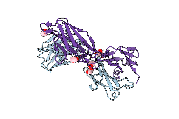 |
Organism: Mus musculus
Method: X-RAY DIFFRACTION Resolution:2.10 Å Release Date: 2020-09-09 Classification: IMMUNE SYSTEM Ligands: EDO |
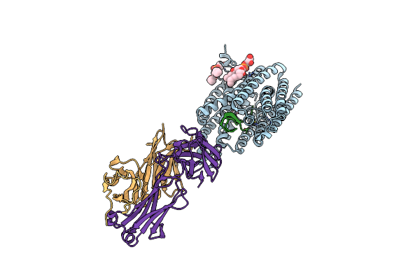 |
Structure Of Human Ferroportin Bound To Hepcidin And Cobalt In Lipid Nanodisc
Organism: Homo sapiens, Mus musculus
Method: ELECTRON MICROSCOPY Release Date: 2020-09-09 Classification: TRANSLOCASE/IMMUNE SYSTEM/HORMONE Ligands: AGA, CO |
 |
Single-Particle Reconstruction Of Darp14 - A Designed Protein Scaffold Displaying ~17Kda Darpin Proteins - Scaffold
|
 |
Single-Particle Reconstruction Of Darp14 - A Designed Protein Scaffold Displaying ~17Kda Darpin Proteins
Organism: Synthetic, Pseudomonas aeruginosa (strain atcc 15692 / dsm 22644 / cip 104116 / jcm 14847 / lmg 12228 / 1c / prs 101 / pao1)
Method: ELECTRON MICROSCOPY Release Date: 2018-03-21 Classification: DE NOVO PROTEIN |
 |
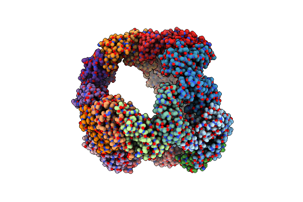
Crystal Structure Of Designed Two-Component Self-Assembling Icosahedral Cage I52-32
Organism: Candida albicans p37005, Caldanaerobacter subterraneus subsp. tengcongensis (strain dsm 15242 / jcm 11007 / nbrc 100824 / mb4)
Method: X-RAY DIFFRACTION Resolution:3.50 Å Release Date: 2016-07-27 Classification: PROTEIN BINDING |
 |
Crystal Structure Of Designed Two-Component Self-Assembling Icosahedral Cage I53-40
Organism: Methanocaldococcus jannaschii (strain atcc 43067 / dsm 2661 / jal-1 / jcm 10045 / nbrc 100440), Vibrionales bacterium (strain swat-3)
Method: X-RAY DIFFRACTION Resolution:3.70 Å Release Date: 2016-07-27 Classification: PROTEIN BINDING |
 |
Crystal Structure Of Designed Two-Component Self-Assembling Icosahedral Cage I32-28
Organism: Burkholderia thailandensis e264, Deinococcus radiodurans r1
Method: X-RAY DIFFRACTION Resolution:5.59 Å Release Date: 2016-07-27 Classification: PROTEIN BINDING |
 |
Crystal Structure Of Rescued Two-Component Self-Assembling Tetrahedral Cage T33-31
Organism: Thermus thermophilus (strain hb8 / atcc 27634 / dsm 579)
Method: X-RAY DIFFRACTION Resolution:3.40 Å Release Date: 2015-07-29 Classification: PROTEIN BINDING |
 |
Computationally Designed Two-Component Self-Assembling Tetrahedral Cage T32-28
Organism: Campylobacter jejuni, Salmonella enterica subsp. enterica serovar typhimurium
Method: X-RAY DIFFRACTION Resolution:4.50 Å Release Date: 2014-05-28 Classification: PROTEIN BINDING |
 |
Computationally Designed Two-Component Self-Assembling Tetrahedral Cage T33-15
Organism: Shewanella oneidensis, Thermus thermophilus
Method: X-RAY DIFFRACTION Resolution:2.80 Å Release Date: 2014-05-28 Classification: PROTEIN BINDING Ligands: CA |
 |
Computationally Designed Two-Component Self-Assembling Tetrahedral Cage, T33-21, Crystallized In Space Group R32
Organism: Pyrococcus horikoshii, Pseudomonas aeruginosa
Method: X-RAY DIFFRACTION Resolution:2.10 Å Release Date: 2014-05-28 Classification: PROTEIN BINDING Ligands: SO4, NH4 |
 |
Computationally Designed Two-Component Self-Assembling Tetrahedral Cage, T33-21, Crystallized In Space Group F4132
Organism: Pyrococcus horikoshii, Pseudomonas aeruginosa
Method: X-RAY DIFFRACTION Resolution:2.80 Å Release Date: 2014-05-28 Classification: PROTEIN BINDING Ligands: SO4 |