
Search Count: 65
 |
Organism: Drosophila melanogaster
Method: ELECTRON MICROSCOPY Release Date: 2023-12-20 Classification: MEMBRANE PROTEIN Ligands: POV, K |
 |
Organism: Escherichia coli k-12
Method: ELECTRON MICROSCOPY Release Date: 2023-02-15 Classification: MEMBRANE PROTEIN |
 |
Organism: Homo sapiens
Method: ELECTRON MICROSCOPY Release Date: 2022-11-23 Classification: TRANSPORT PROTEIN Ligands: K, 4AP |
 |
Organism: Homo sapiens
Method: ELECTRON MICROSCOPY Release Date: 2022-06-29 Classification: MEMBRANE PROTEIN Ligands: K |
 |
Organism: Homo sapiens
Method: ELECTRON MICROSCOPY Release Date: 2022-06-01 Classification: MEMBRANE PROTEIN Ligands: K |
 |
Organism: Drosophila melanogaster
Method: ELECTRON MICROSCOPY Release Date: 2022-03-30 Classification: MEMBRANE PROTEIN Ligands: POV, K |
 |
Organism: Drosophila melanogaster
Method: ELECTRON MICROSCOPY Release Date: 2022-03-30 Classification: MEMBRANE PROTEIN Ligands: POV, K |
 |
Organism: Homo sapiens
Method: X-RAY DIFFRACTION Resolution:2.88 Å Release Date: 2022-02-02 Classification: MEMBRANE PROTEIN,TRANSFERASE Ligands: ZN, PKZ, PO4 |
 |
The Structure Of Bacillus Subtilis Bmrcd In The Inward-Facing Conformation Bound To Hoechst-33342 And Atp
Organism: Bacillus subtilis subsp. subtilis str. 168
Method: ELECTRON MICROSCOPY Release Date: 2022-01-05 Classification: TRANSPORT PROTEIN Ligands: ATP, HT1 |
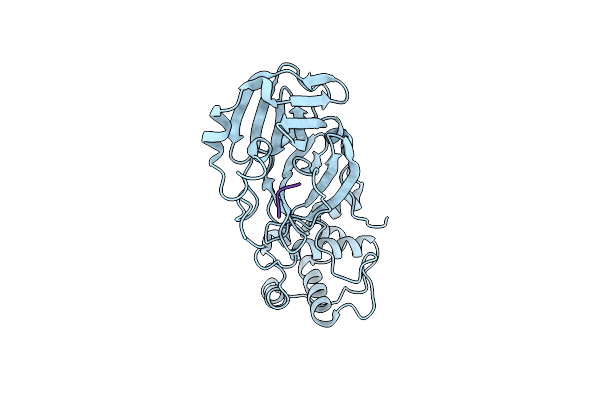 |
Organism: Severe acute respiratory syndrome coronavirus 2, Synthetic construct
Method: X-RAY DIFFRACTION Resolution:1.70 Å Release Date: 2021-08-25 Classification: Viral protein/hydrolase inhibitor |
 |
Monomer Yeast Atp Synthase Fo Reconstituted In Nanodisc With Inhibitor Of Bedaquiline Bound
Organism: Saccharomyces cerevisiae, Saccharomyces cerevisiae (strain atcc 204508 / s288c)
Method: ELECTRON MICROSCOPY Release Date: 2020-08-26 Classification: BIOSYNTHETIC PROTEIN |
 |
Lmrp From L. Lactis, In An Outward-Open Conformation, Bound To Hoechst 33342
Organism: Lactococcus lactis
Method: X-RAY DIFFRACTION Resolution:2.90 Å Release Date: 2020-07-29 Classification: TRANSPORT PROTEIN Ligands: HT1, XP4, LMU, GOL |
 |
Structure Of Human Atg9A, The Only Transmembrane Protein Of The Core Autophagy Machinery
Organism: Homo sapiens
Method: ELECTRON MICROSCOPY Release Date: 2020-07-08 Classification: MEMBRANE PROTEIN Ligands: LMN |
 |
Structure Of Human Atg9A, The Only Transmembrane Protein Of The Core Autophagy Machinery
Organism: Homo sapiens
Method: ELECTRON MICROSCOPY Release Date: 2020-07-08 Classification: MEMBRANE PROTEIN Ligands: LMN |
 |
Organism: Homo sapiens
Method: X-RAY DIFFRACTION Resolution:3.30 Å Release Date: 2019-06-12 Classification: MEMBRANE PROTEIN Ligands: ATP, NAG, EDO, CA |
 |
Organism: Homo sapiens
Method: X-RAY DIFFRACTION Resolution:3.82 Å Release Date: 2019-06-12 Classification: MEMBRANE PROTEIN Ligands: ATP, NAG, EDO, MG |
 |
Monomer Yeast Atp Synthase (F1Fo) Reconstituted In Nanodisc With Inhibitor Of Oligomycin Bound.
Organism: Saccharomyces cerevisiae, Saccharomyces cerevisiae (strain atcc 204508 / s288c)
Method: ELECTRON MICROSCOPY Release Date: 2018-04-11 Classification: BIOSYNTHETIC PROTEIN Ligands: ATP, ADP |
 |
Monomer Yeast Atp Synthase Fo Reconstituted In Nanodisc With Inhibitor Of Oligomycin Bound Generated From Focused Refinement.
Organism: Saccharomyces cerevisiae (strain atcc 204508 / s288c)
Method: ELECTRON MICROSCOPY Release Date: 2018-04-11 Classification: BIOSYNTHETIC PROTEIN Ligands: EFO |
 |
Organism: Saccharomyces cerevisiae, Saccharomyces cerevisiae (strain atcc 204508 / s288c)
Method: ELECTRON MICROSCOPY Release Date: 2018-04-11 Classification: BIOSYNTHETIC PROTEIN Ligands: ATP, ADP |
 |
Monomer Yeast Atp Synthase Fo Reconstituted In Nanodisc Generated From Masked Refinement.
Organism: Saccharomyces cerevisiae (strain atcc 204508 / s288c)
Method: ELECTRON MICROSCOPY Release Date: 2018-04-11 Classification: BIOSYNTHETIC PROTEIN |