
Search Count: 28
 |
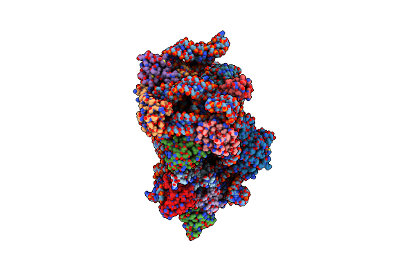
Organism: Ranoidea citropa
Method: X-RAY DIFFRACTION Release Date: 2025-10-08 Classification: ANTIMICROBIAL PROTEIN |
 |
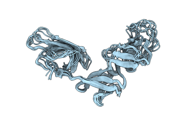
Amyloid Fibril From The Antimicrobial Peptide Citropin 1-3 - Polymorph Nr.3
Organism: Ranoidea citropa
Method: ELECTRON MICROSCOPY Release Date: 2025-10-01 Classification: ANTIMICROBIAL PROTEIN |
 |
Amyloid Fibril From The Antimicrobial Peptide Citropin 1-3 - Polymorph Nr.2
Organism: Ranoidea citropa
Method: ELECTRON MICROSCOPY Release Date: 2025-10-01 Classification: ANTIMICROBIAL PROTEIN |
 |
Amyloid Fibril From The Antimicrobial Peptide Citropin 1-3 - Polymorph Nr.1L
Organism: Ranoidea citropa
Method: ELECTRON MICROSCOPY Release Date: 2025-10-01 Classification: ANTIMICROBIAL PROTEIN |
 |
Amyloid Fibril From The Antimicrobial Peptide Citropin 1-3 - Polymorph Nr.3L
Organism: Ranoidea citropa
Method: ELECTRON MICROSCOPY Release Date: 2025-10-01 Classification: ANTIMICROBIAL PROTEIN |
 |
Amyloid Fibril From The Antimicrobial Peptide Citropin 1-3 - Polymorph Nr.2L
Organism: Ranoidea citropa
Method: ELECTRON MICROSCOPY Release Date: 2025-10-01 Classification: ANTIMICROBIAL PROTEIN |
 |
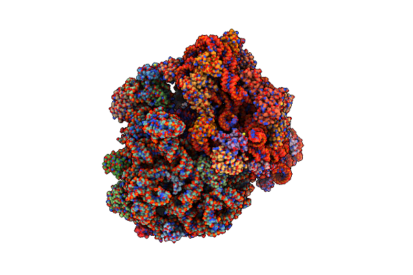
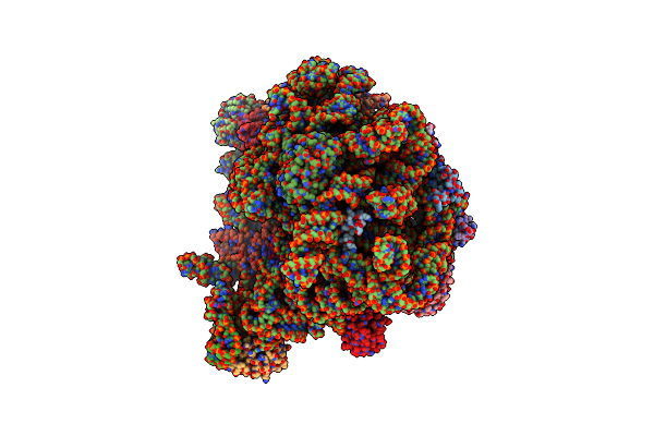
Organism: Staphylococcus aureus subsp. aureus nctc 8325
Method: ELECTRON MICROSCOPY Release Date: 2023-12-27 Classification: RIBOSOME |
 |
Organism: Staphylococcus aureus subsp. aureus nctc 8325
Method: X-RAY DIFFRACTION Resolution:2.22 Å Release Date: 2023-02-22 Classification: RIBOSOME |
 |
70S Initiation Complex With Assigned Rrna Modifications From Staphylococcus Aureus
Organism: Synthetic construct, Staphylococcus aureus subsp. aureus nctc 8325, Staphylococcus aureus (strain nctc 8325), Escherichia coli dh5[alpha]
Method: ELECTRON MICROSCOPY Release Date: 2021-01-27 Classification: RIBOSOME Ligands: MG, K |
 |
Organism: Staphylococcus aureus subsp. aureus, Staphylococcus aureus subsp. aureus nctc 8325
Method: X-RAY DIFFRACTION Resolution:2.27 Å Release Date: 2020-04-15 Classification: RIBOSOME Ligands: ACY, PO4 |
 |
Organism: Staphylococcus aureus (strain nctc 8325)
Method: ELECTRON MICROSCOPY Release Date: 2020-04-08 Classification: RIBOSOME Ligands: MG, K |
 |
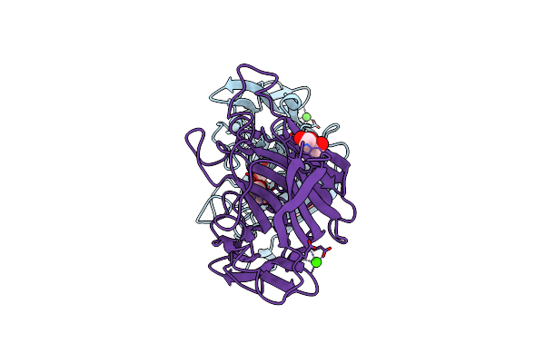
Organism: Staphylococcus aureus (strain nctc 8325)
Method: X-RAY DIFFRACTION Resolution:1.48 Å Release Date: 2020-04-01 Classification: TRANSLATION |
 |
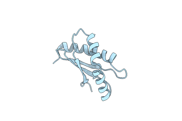
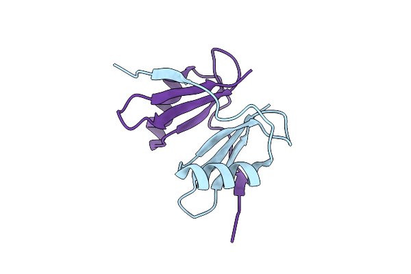
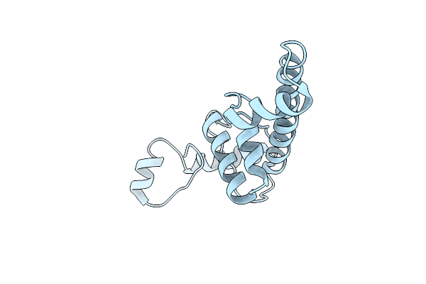
Solution Structure Of The Ribosome Elongation Factor P (Ef-P) From Staphylococcus Aureus
Organism: Staphylococcus aureus
Method: SOLUTION NMR Release Date: 2020-03-11 Classification: STRUCTURAL PROTEIN |
 |
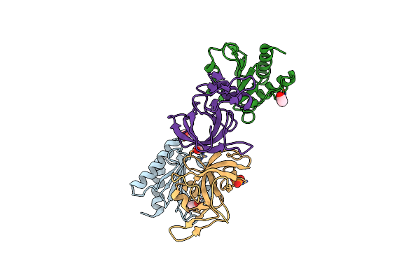
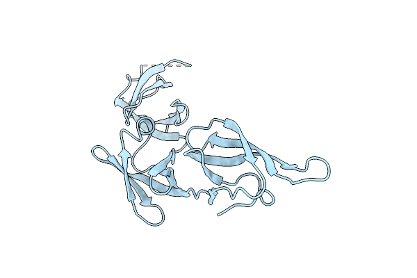
X-Ray Structure Of The C-Terminal Domain Of S. Aureus Hibernating Promoting Factor (Ctd-Sahpf)
Organism: Staphylococcus aureus (strain nctc 8325)
Method: X-RAY DIFFRACTION Resolution:1.60 Å Release Date: 2019-12-11 Classification: RIBOSOMAL PROTEIN |
 |
Organism: Xylella fastidiosa (strain 9a5c)
Method: X-RAY DIFFRACTION Resolution:3.00 Å Release Date: 2016-01-27 Classification: DNA BINDING PROTEIN |
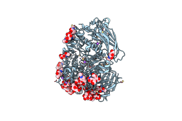 |
Structure Of Fungal Beta-Mannosidase From Glycoside Hydrolase Family 2 Of Trichoderma Harzianum
Organism: Trichoderma harzianum
Method: X-RAY DIFFRACTION Resolution:1.90 Å Release Date: 2014-07-09 Classification: HYDROLASE Ligands: NAG, CD, NA, PG4, PEG, CA, CL |
 |
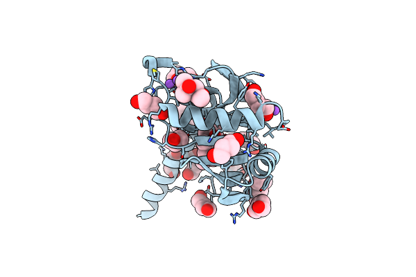
Organism: Hypocrea lixii
Method: X-RAY DIFFRACTION Resolution:2.50 Å Release Date: 2014-07-09 Classification: HYDROLASE Ligands: NAG, CD, NA, 1PE, PE4, CL, CA |
 |
Crystal Structure Of A Putative Nucleotidyltransferase (Tm1012) From Thermotoga Maritima At 1.87 A Resolution
Organism: Thermotoga maritima
Method: X-RAY DIFFRACTION Resolution:1.87 Å Release Date: 2013-09-11 Classification: TRANSFERASE |
 |
Organism: Rhodothermus marinus
Method: X-RAY DIFFRACTION Resolution:1.95 Å Release Date: 2010-08-18 Classification: HYDROLASE Ligands: GOL, CA |
 |
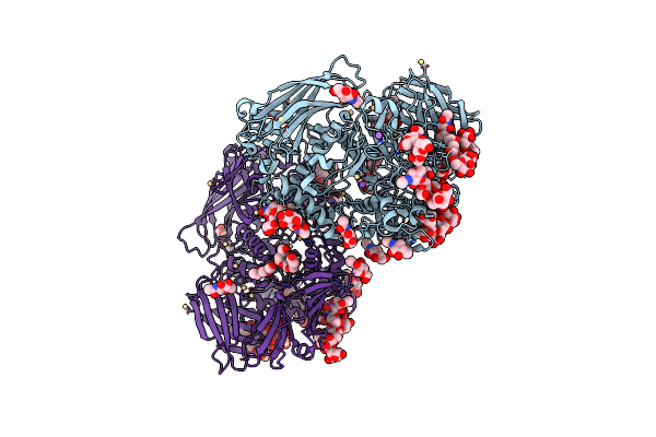
Crystal Structure Of Trichoderma Reesei Aspartic Proteinase Complexed With Pepstatin A
Organism: Streptomyces argenteolus subsp. toyonakensis, Hypocrea jecorina
Method: X-RAY DIFFRACTION Resolution:1.85 Å Release Date: 2008-10-07 Classification: HYDROLASE/HYDROLASE INHIBITOR |