Search Count: 12
 |
Organism: Pseudomonas putida
Method: X-RAY DIFFRACTION Resolution:2.10 Å Release Date: 2011-03-23 Classification: LYASE Ligands: 3CO, GOL |
 |
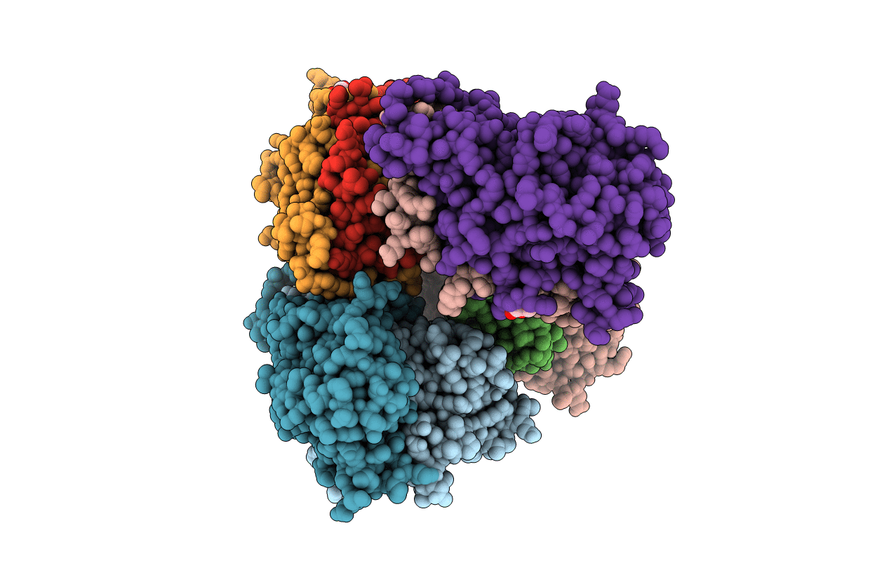
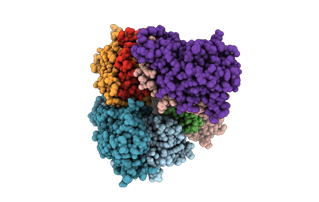
Crystal Structure Of Co-Type Nitrile Hydratase Beta-E56Q From Pseudomonas Putida.
Organism: Pseudomonas putida
Method: X-RAY DIFFRACTION Resolution:2.30 Å Release Date: 2011-03-23 Classification: LYASE Ligands: GOL, 3CO |
 |
Crystal Structure Of Co-Type Nitrile Hydratase Beta-H71L From Pseudomonas Putida.
Organism: Pseudomonas putida
Method: X-RAY DIFFRACTION Resolution:2.00 Å Release Date: 2011-03-23 Classification: LYASE Ligands: 3CO |
 |
Crystal Structure Of Co-Type Nitrile Hydratase Alpha-E168Q From Pseudomonas Putida.
Organism: Pseudomonas putida
Method: X-RAY DIFFRACTION Resolution:2.50 Å Release Date: 2011-03-23 Classification: LYASE Ligands: GOL, 3CO |
 |
Crystal Structure Of Co-Type Nitrile Hydratase Beta-Y215F From Pseudomonas Putida.
Organism: Pseudomonas putida
Method: X-RAY DIFFRACTION Resolution:2.40 Å Release Date: 2011-03-23 Classification: LYASE Ligands: GOL, 3CO |
 |
Solution Structure Of A Designed Spirocyclic Helical Ligand Binding At A Two-Base Bulge Site In Dna
|
 |
Solution Structure Of The Complex Formed Between A Left-Handed Wedge-Shaped Spirocyclic Molecule And Bulged Dna
|
 |
Solution Structure Of A Wedge-Shaped Synthetic Molecule At A Two-Base Bulge Site In Dna
|
 |
A Third Complex Of Post-Activated Neocarzinostatin Chromophore With Dna. Bulge Dna Binding From The Minor Groove
|
 |
Ncsi-Gb-Bulge-Dna Complex Induced Formation Of A Dna Bulge Structure By A Molecular Wedge Ligand-Post-Activated Neocarzinostatin Chromophore
|
 |
Organism: Human papillomavirus type 16
Method: X-RAY DIFFRACTION Resolution:3.50 Å Release Date: 2000-03-09 Classification: VIRUS |
 |
Solution Nmr Structure Of A Two-Base Dna Bulge Complexed With An Enediyne Cleaving Analog, 11 Structures
|

