
Search Count: 18
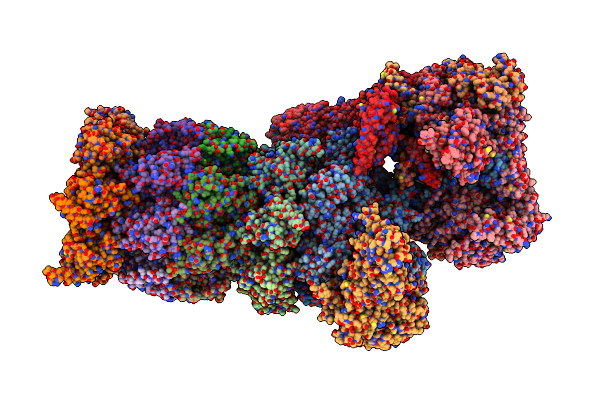 |
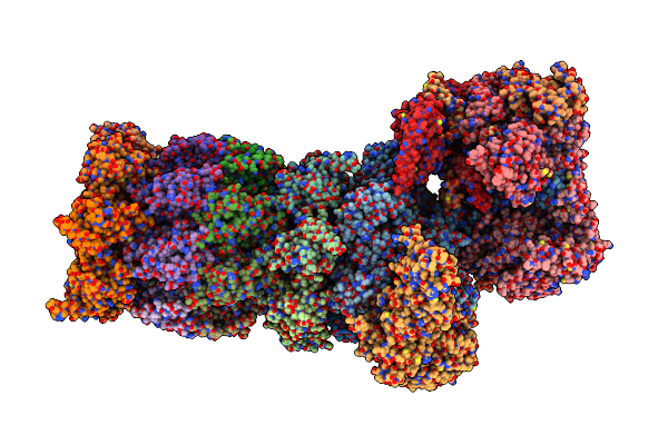
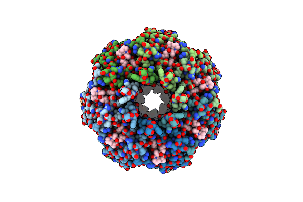
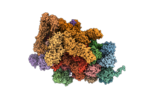
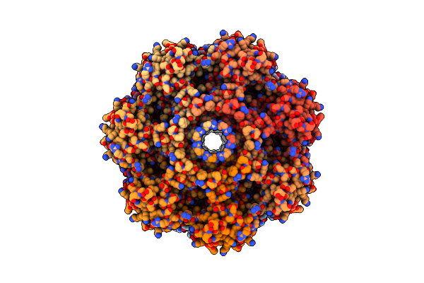
Organism: Homo sapiens
Method: ELECTRON MICROSCOPY Release Date: 2023-02-08 Classification: STRUCTURAL PROTEIN Ligands: ZN, ATP, MG, ADP |
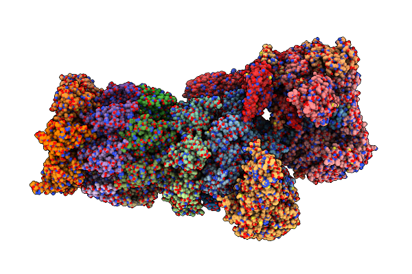 |
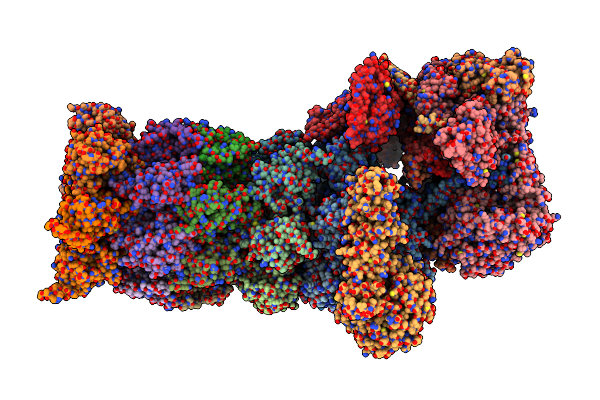
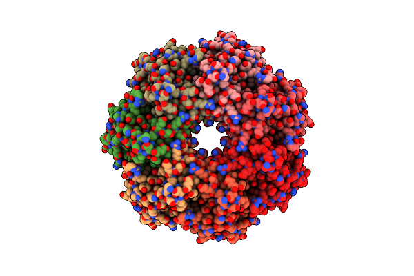
Organism: Homo sapiens
Method: ELECTRON MICROSCOPY Release Date: 2023-02-08 Classification: STRUCTURAL PROTEIN Ligands: ZN, ATP, MG, ADP |
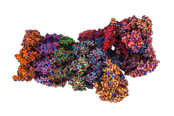 |
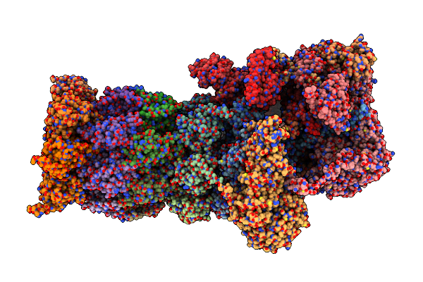
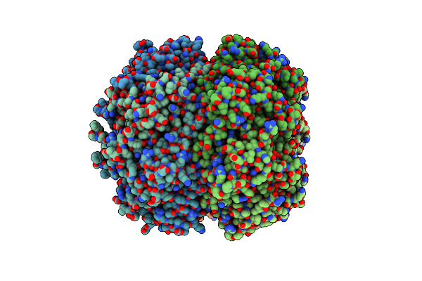
Organism: Homo sapiens
Method: ELECTRON MICROSCOPY Release Date: 2023-02-08 Classification: STRUCTURAL PROTEIN Ligands: ZN, ATP, MG, ADP |
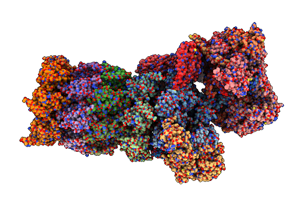 |
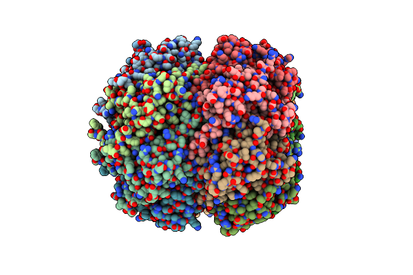
Organism: Homo sapiens
Method: ELECTRON MICROSCOPY Release Date: 2023-02-08 Classification: STRUCTURAL PROTEIN Ligands: ZN, ATP, MG, ADP |
 |
Organism: Homo sapiens
Method: ELECTRON MICROSCOPY Release Date: 2023-02-08 Classification: STRUCTURAL PROTEIN Ligands: ZN, ATP, MG, ADP |
 |
Organism: Homo sapiens
Method: ELECTRON MICROSCOPY Release Date: 2023-02-08 Classification: STRUCTURAL PROTEIN Ligands: ZN, ATP |
 |
Organism: Homo sapiens
Method: ELECTRON MICROSCOPY Release Date: 2023-02-08 Classification: STRUCTURAL PROTEIN Ligands: ZN, ATP |
 |
Organism: Homo sapiens
Method: ELECTRON MICROSCOPY Release Date: 2023-02-08 Classification: STRUCTURAL PROTEIN Ligands: ZN, ATP, MG, ADP |
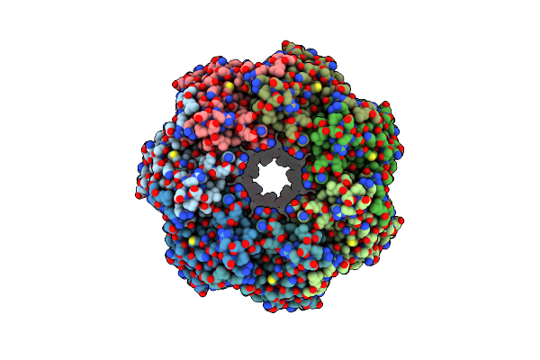 |
Organism: Mycobacterium tuberculosis
Method: ELECTRON MICROSCOPY Release Date: 2020-03-18 Classification: HYDROLASE |
 |
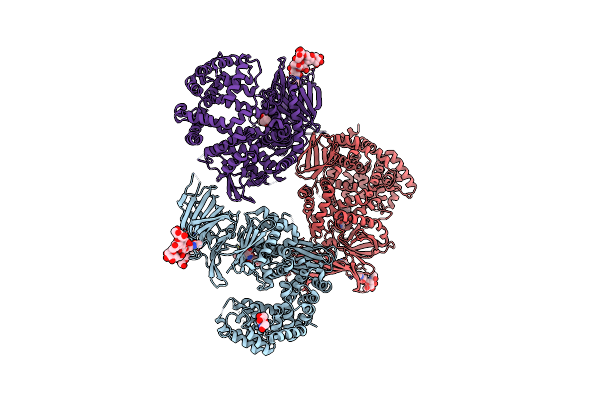
Organism: Mycobacterium tuberculosis, Synthetic construct
Method: ELECTRON MICROSCOPY Release Date: 2020-03-18 Classification: HYDROLASE/ANTIBIOTIC |
 |
Organism: Mycobacterium tuberculosis, Synthetic construct
Method: ELECTRON MICROSCOPY Release Date: 2020-03-18 Classification: HYDROLASE |
 |
Crystal Structure Of The Active Form Of The Proteolytic Complex Clpp1 And Clpp2
Organism: Mycobacterium tuberculosis (strain cdc 1551 / oshkosh), Synthetic construct
Method: X-RAY DIFFRACTION Resolution:3.07 Å Release Date: 2016-02-17 Classification: HYDROLASE |
 |
Crystal Structure Of The Active Form Of The Proteolytic Complex Clpp1 And Clpp2
Organism: Mycobacterium tuberculosis (strain cdc 1551 / oshkosh)
Method: X-RAY DIFFRACTION Resolution:2.90 Å Release Date: 2016-02-17 Classification: HYDROLASE |
 |
Organism: Saccharomyces cerevisiae, Homo sapiens
Method: ELECTRON MICROSCOPY Resolution:9.50 Å Release Date: 2015-07-22 Classification: HYDROLASE |
 |
Er Aminopeptidase, Erap1, Bound To The Zinc Aminopeptidase Inhibitor, Bestatin
Organism: Homo sapiens
Method: X-RAY DIFFRACTION Resolution:2.95 Å Release Date: 2011-03-30 Classification: Hydrolase/Hydrolase Inhibitor Ligands: ZN, BES, NAG |
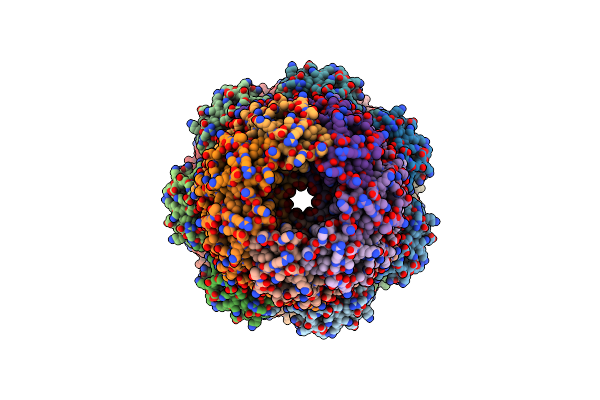 |
Crystal Structure Of Archaeal 20S Proteasome In Complex With The C-Terminus Of Pan
Organism: Thermoplasma acidophilum, Trypanosoma brucei brucei, Methanocaldococcus jannaschii
Method: X-RAY DIFFRACTION Resolution:4.00 Å Release Date: 2009-12-29 Classification: HYDROLASE/HYDROLASE ACTIVATOR |
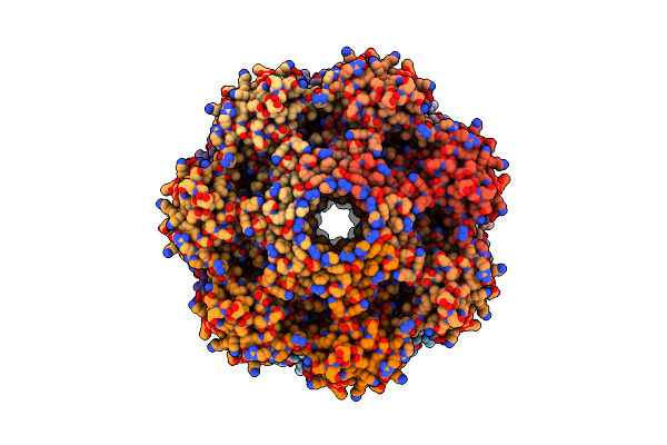 |
Organism: Thermoplasma acidophilum
Method: ELECTRON MICROSCOPY Resolution:6.80 Å Release Date: 2008-08-05 Classification: HYDROLASE |
 |
Organism: Thermoplasma acidophilum
Method: ELECTRON MICROSCOPY Resolution:6.80 Å Release Date: 2008-08-05 Classification: HYDROLASE |