
Search Count: 40
 |
Organism: Caenorhabditis elegans
Method: ELECTRON MICROSCOPY Release Date: 2024-03-27 Classification: MEMBRANE PROTEIN Ligands: TWT, PEE, R16, CLR, NAG, CA, PLM |
 |
Organism: Caenorhabditis elegans
Method: ELECTRON MICROSCOPY Release Date: 2022-10-19 Classification: MEMBRANE PROTEIN Ligands: CA, PEE, 3PE, ZFC, D12, CLR, R16, NAG, PLM |
 |
Organism: Caenorhabditis elegans
Method: ELECTRON MICROSCOPY Release Date: 2022-10-19 Classification: MEMBRANE PROTEIN Ligands: CA, PEE, D12, R16, CLR, NAG, 3PE, PLM |
 |
Organism: Caenorhabditis elegans
Method: ELECTRON MICROSCOPY Release Date: 2022-10-19 Classification: MEMBRANE PROTEIN Ligands: CA, PEE, 3PE, NAG, PLM |
 |
Organism: Trypanosoma brucei brucei (strain 927/4 gutat10.1)
Method: X-RAY DIFFRACTION Resolution:1.35 Å Release Date: 2019-03-20 Classification: ISOMERASE |
 |
Organism: Mus musculus
Method: X-RAY DIFFRACTION Resolution:1.40 Å Release Date: 2018-08-15 Classification: membrane protein, metal transport Ligands: MAN |
 |
Organism: Mus musculus
Method: ELECTRON MICROSCOPY Release Date: 2018-08-15 Classification: MEMBRANE PROTEIN |
 |
Organism: Mus musculus
Method: ELECTRON MICROSCOPY Release Date: 2018-08-15 Classification: membrane protein, metal transport |
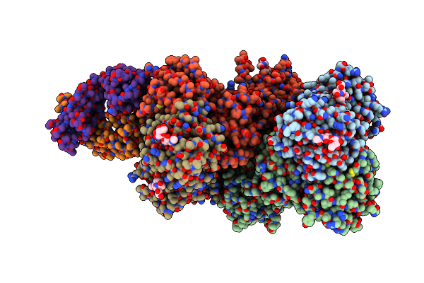 |
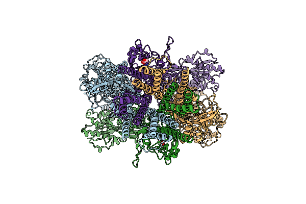
Triheteromeric Nmda Receptor Glun1/Glun2A/Glun2B In Complex With Glycine, Glutamate, Mk-801 And A Glun2B-Specific Fab, At Ph 6.5
Organism: Xenopus laevis, Mus musculus
Method: ELECTRON MICROSCOPY Resolution:4.50 Å Release Date: 2017-03-22 Classification: MEMBRANE PROTEIN Ligands: GLU, BMK |
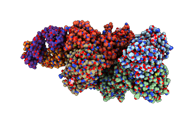 |
Triheteromeric Nmda Receptor Glun1/Glun2A/Glun2B In Complex With Glycine, Glutamate, Ro 25-6981, Mk-801 And A Glun2B-Specific Fab, At Ph 6.5
Organism: Xenopus laevis, Mus musculus
Method: ELECTRON MICROSCOPY Release Date: 2017-03-22 Classification: MEMBRANE PROTEIN Ligands: NAG |
 |
Organism: Trypanosoma brucei brucei treu927
Method: SOLUTION NMR Release Date: 2016-10-12 Classification: ISOMERASE |
 |
Organism: Trypanosoma brucei brucei treu927
Method: SOLUTION NMR Release Date: 2016-10-12 Classification: ISOMERASE |
 |
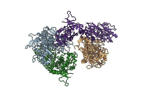
Cryo-Em Structure Of Glun1/Glun2B Nmda Receptor In The Glutamate/Glycine-Bound Conformation
Organism: Xenopus laevis
Method: ELECTRON MICROSCOPY Resolution:7.00 Å Release Date: 2016-04-20 Classification: SIGNALING PROTEIN Ligands: GLY, GLU |
 |
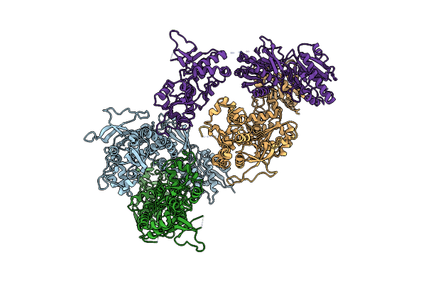
Cryo-Em Structure Of Glun1/Glun2B Nmda Receptor In The Glutamate/Glycine/Ro25-6981-Bound Conformation
Organism: Xenopus laevis
Method: ELECTRON MICROSCOPY Resolution:7.50 Å Release Date: 2016-04-20 Classification: SIGNALING PROTEIN Ligands: GLY, QEM, GLU |
 |
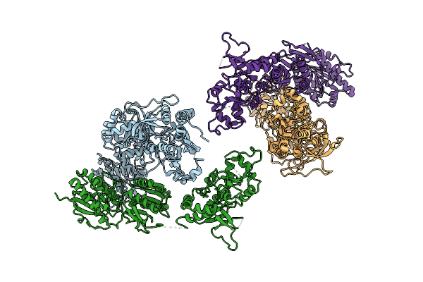
Cryo-Em Structure Of Glun1/Glun2B Nmda Receptor In The Dcka/D-Apv-Bound Conformation, State 2
Organism: Xenopus laevis
Method: ELECTRON MICROSCOPY Release Date: 2016-04-20 Classification: SIGNALING PROTEIN |
 |
Cryo-Em Structure Of Glun1/Glun2B Nmda Receptor In The Dcka/D-Apv-Bound Conformation, State 3
Organism: Xenopus laevis
Method: ELECTRON MICROSCOPY Release Date: 2016-04-20 Classification: SIGNALING PROTEIN |
 |
Cryo-Em Structure Of Glun1/Glun2B Nmda Receptor In The Dcka/D-Apv-Bound Conformation, State 4
Organism: Xenopus laevis
Method: ELECTRON MICROSCOPY Release Date: 2016-04-20 Classification: SIGNALING PROTEIN |
 |
Cryo-Em Structure Of Glun1/Glun2B Nmda Receptor In The Dcka/D-Apv-Bound Conformation, State 5
Organism: Xenopus laevis
Method: ELECTRON MICROSCOPY Release Date: 2016-04-20 Classification: SIGNALING PROTEIN |
 |
Cryo-Em Structure Of Glun1/Glun2B Nmda Receptor In The Dcka/D-Apv-Bound Conformation, State 6
Organism: Xenopus laevis
Method: ELECTRON MICROSCOPY Release Date: 2016-04-20 Classification: SIGNALING PROTEIN |
 |
Cryo-Em Structure Of Glun1/Glun2B Nmda Receptor In The Dcka/D-Apv-Bound Conformation, State 1
Organism: Xenopus laevis
Method: ELECTRON MICROSCOPY Release Date: 2016-04-20 Classification: SIGNALING PROTEIN |