
Search Count: 4
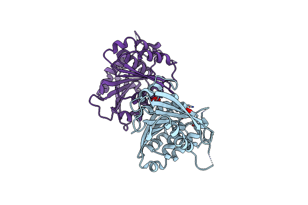 |
Crystal Structure Of The Ferric Enterobactin Esterase (Pfee) From Pseudomonas Aeruginosa
Organism: Pseudomonas aeruginosa
Method: X-RAY DIFFRACTION Resolution:2.00 Å Release Date: 2018-06-20 Classification: HYDROLASE Ligands: NO3 |
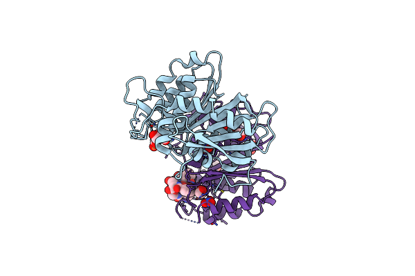 |
Crystal Structure Of The Ferric Enterobactin Esterase (Pfee) Mutant(S157A) From Pseudomonas Aeruginosa In Presence Of Enterobactin
Organism: Pseudomonas aeruginosa pao1
Method: X-RAY DIFFRACTION Resolution:1.66 Å Release Date: 2018-06-20 Classification: HYDROLASE Ligands: FE, DBS, NO3, EDO, EB4 |
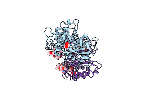 |
Crystal Structure Of The Ferric Enterobactin Esterase (Pfee) Mutant(S157A) From Pseudomonas Aeruginosa In Complex With Tris-Catechol Vector
Organism: Pseudomonas aeruginosa
Method: X-RAY DIFFRACTION Resolution:1.49 Å Release Date: 2018-06-20 Classification: HYDROLASE Ligands: FE, 8SW, NO3, EDO |
 |
Crystal Structure Of The Ferric Enterobactin Esterase (Pfee) From Pseudomonas Aeruginosa In Complex With The Tris-Catechol Vector
Organism: Pseudomonas aeruginosa
Method: X-RAY DIFFRACTION Resolution:3.11 Å Release Date: 2018-06-20 Classification: HYDROLASE Ligands: FE, 8SW |