
Search Count: 88
 |
Organism: Synthetic construct
Method: ELECTRON MICROSCOPY Release Date: 2025-12-10 Classification: CHAPERONE Ligands: ADP, MG |
 |
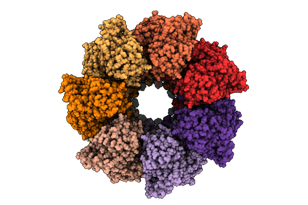
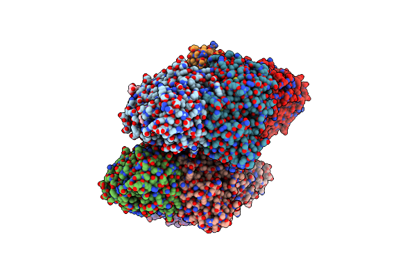
Ancestral Group Iii Chaperonin (Aciii) Double-Ring In Open Conformation Including The Equatorial And Intermediate Domains (Residues 12-200 And 356-507)
Organism: Synthetic construct
Method: ELECTRON MICROSCOPY Release Date: 2025-12-10 Classification: CHAPERONE |
 |
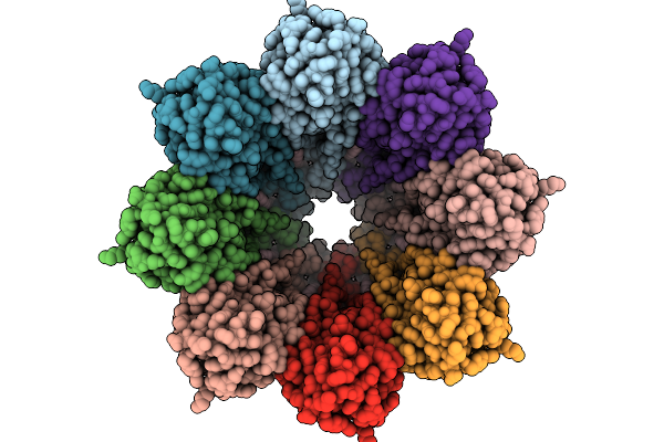
Ancestral Fibrobacteres-Chlorobi-Bacteroidetes Group Chaperonin (Afcb) Double-Ring In Open Conformation
Organism: Synthetic construct
Method: ELECTRON MICROSCOPY Release Date: 2025-12-10 Classification: CHAPERONE |
 |
Organism: Stichodactyla helianthus
Method: ELECTRON MICROSCOPY Release Date: 2025-10-29 Classification: MEMBRANE PROTEIN Ligands: FO4, CLR |
 |
Organism: Actinia fragacea
Method: ELECTRON MICROSCOPY Release Date: 2025-10-29 Classification: MEMBRANE PROTEIN Ligands: FO4, CLR |
 |
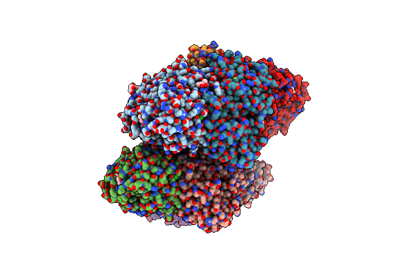
Structure Of The Octameric Pore Of Fragaceotxin C (Frac Or Delta-Actitoxin-Afr1A) In Large Unilamellar Vesicles.
Organism: Actinia fragacea
Method: ELECTRON MICROSCOPY Release Date: 2025-10-08 Classification: MEMBRANE PROTEIN Ligands: FO4, CLR |
 |
Structure Of 6Mer Pore Intermediate Of Sticholysin Ii (Stnii) Toxin In Lipid Nanodiscs
Organism: Stichodactyla helianthus
Method: ELECTRON MICROSCOPY Release Date: 2025-10-08 Classification: MEMBRANE PROTEIN Ligands: FO4 |
 |
Organism: Homo sapiens
Method: X-RAY DIFFRACTION Release Date: 2025-10-08 Classification: IMMUNE SYSTEM Ligands: GOL, SO4 |
 |
Type-I Interferons Autoantibodies Pmab15 And Pmab14 In Complex With Interferon Alpha-2
Organism: Homo sapiens
Method: X-RAY DIFFRACTION Release Date: 2025-10-08 Classification: IMMUNE SYSTEM Ligands: GOL, PO4, K |
 |
Type-I Interferon Autoantibodies Pmab3, Pmab19 And Pmab14 In Complex With Interferon Alpha-2
Organism: Homo sapiens
Method: X-RAY DIFFRACTION Release Date: 2025-10-08 Classification: IMMUNE SYSTEM |
 |
Structure Of 5Mer Pore Intermediate Of Sticholysin Ii (Stnii) Toxin In Lipid Nanodiscs
Organism: Stichodactyla helianthus
Method: ELECTRON MICROSCOPY Release Date: 2025-09-17 Classification: MEMBRANE PROTEIN Ligands: FO4 |
 |
Organism: Legionella pneumophila str. corby
Method: X-RAY DIFFRACTION Release Date: 2025-06-18 Classification: HYDROLASE Ligands: A1IRK, ZN |
 |
Organism: Legionella pneumophila str. corby
Method: X-RAY DIFFRACTION Release Date: 2025-06-18 Classification: HYDROLASE Ligands: A1IRL, BU3, GOL, ZN, SO4 |
 |
Organism: Rhodococcus jostii rha1
Method: X-RAY DIFFRACTION Resolution:1.47 Å Release Date: 2025-01-29 Classification: OXIDOREDUCTASE Ligands: FAD, B3P, MPD, MRD, CL |
 |
Organism: Rhodococcus jostii rha1
Method: X-RAY DIFFRACTION Resolution:2.76 Å Release Date: 2024-12-18 Classification: OXIDOREDUCTASE Ligands: FAD |
 |
Organism: Rhodococcus jostii rha1
Method: X-RAY DIFFRACTION Resolution:1.60 Å Release Date: 2024-12-18 Classification: OXIDOREDUCTASE Ligands: FAD, B3P, MPD, CL |
 |
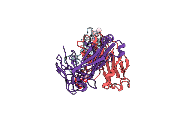
Crystal Structure Of Plasmodium Vivax (Palo Alto) Pvama1 In Complex With Human Fab 826827
Organism: Plasmodium vivax, Homo sapiens
Method: X-RAY DIFFRACTION Resolution:2.40 Å Release Date: 2024-11-13 Classification: CELL INVASION |
 |
Organism: Pectobacterium phage zf40
Method: ELECTRON MICROSCOPY Release Date: 2024-07-24 Classification: RNA BINDING PROTEIN |
 |
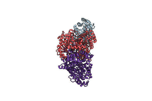
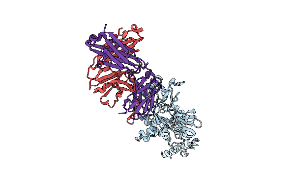
Sars-Cov-2 Spike In Complex With The 17T2 Neutralizing Antibody Fab Fragment (Local Refinement Of Rbd And Fab)
Organism: Severe acute respiratory syndrome coronavirus, Homo sapiens
Method: ELECTRON MICROSCOPY Release Date: 2024-01-10 Classification: VIRAL PROTEIN |
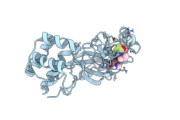 |
Organism: Severe acute respiratory syndrome coronavirus 2
Method: X-RAY DIFFRACTION Resolution:2.00 Å Release Date: 2023-08-30 Classification: VIRAL PROTEIN, Hydrolase/Inhibitor Ligands: YVZ |