
Search Count: 18
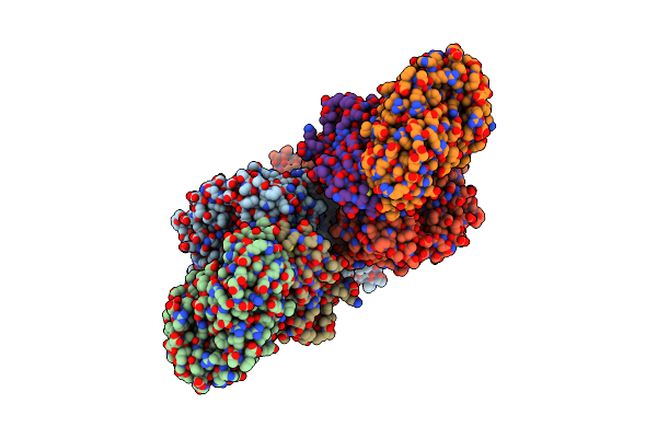 |
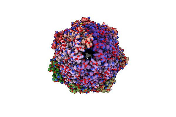
The Cryoem Structure Of The High Affinity Carbon Monoxide Dehydrogenase From Mycobacterium Smegmatis
Organism: Mycolicibacterium smegmatis mc2 155
Method: ELECTRON MICROSCOPY Release Date: 2024-10-16 Classification: OXIDOREDUCTASE Ligands: CUN, MCN, FAD, FES |
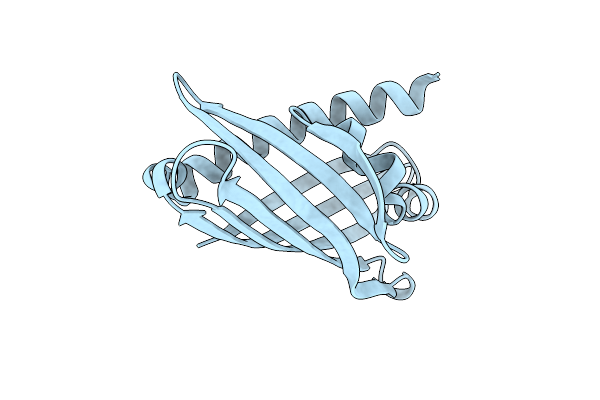 |
The Crystal Structure Of Coxg From M. Smegmatis, Minus Lipid Anchoring C-Terminus.
Organism: Mycolicibacterium smegmatis mc2 155
Method: X-RAY DIFFRACTION Release Date: 2024-10-02 Classification: ELECTRON TRANSPORT |
 |
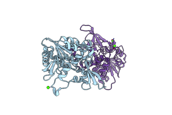
Organism: Plasmodium falciparum (isolate 3d7)
Method: X-RAY DIFFRACTION Resolution:1.80 Å Release Date: 2022-06-08 Classification: LIGASE Ligands: 69X, MLI, CL, MG |
 |
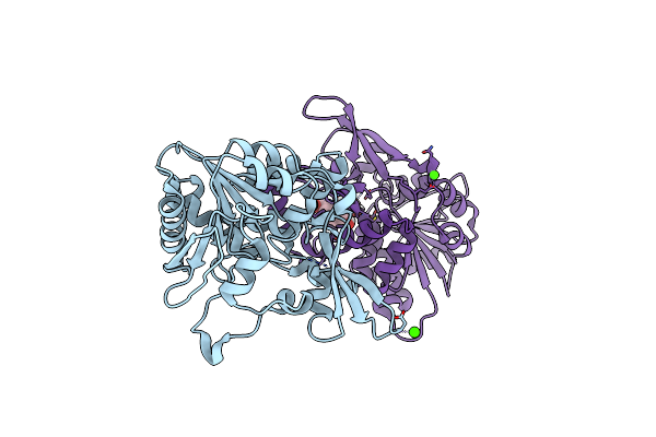
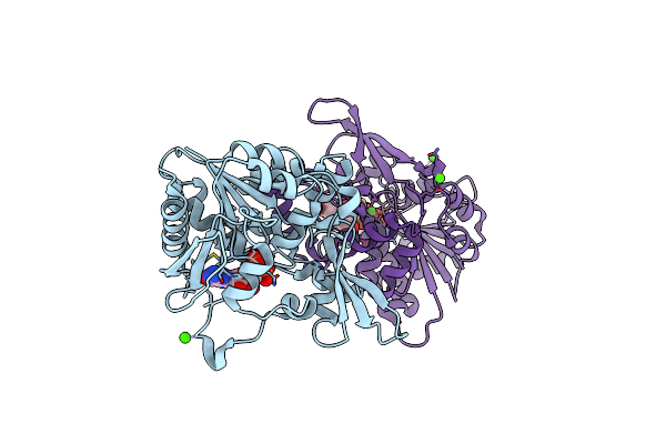
Organism: Plasmodium falciparum (isolate 3d7)
Method: X-RAY DIFFRACTION Resolution:2.15 Å Release Date: 2022-06-08 Classification: LIGASE Ligands: 66I, MG, CL |
 |
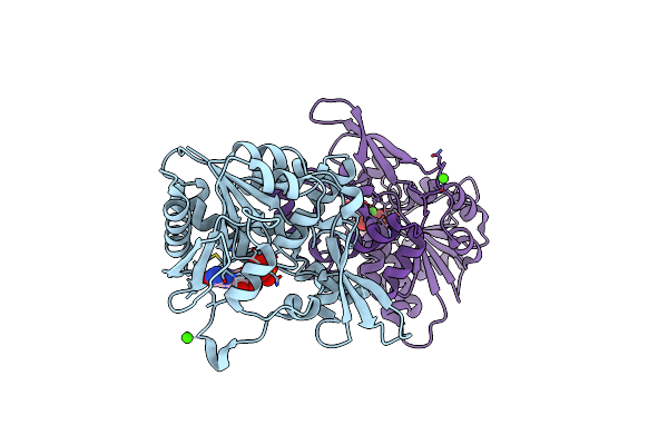
Plasmodium Falciparum Tyrosyl-Trna Synthetase, S234C Mutant, In Complex With Ml901-Tyr
Organism: Plasmodium falciparum (isolate 3d7)
Method: X-RAY DIFFRACTION Resolution:2.20 Å Release Date: 2022-06-08 Classification: LIGASE Ligands: 66I, MG, CL |
 |
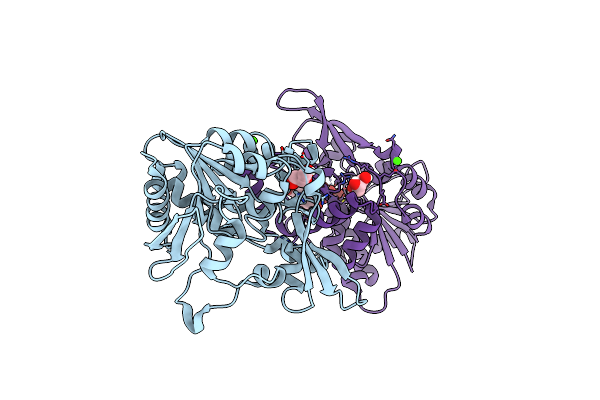
Organism: Homo sapiens
Method: X-RAY DIFFRACTION Resolution:1.70 Å Release Date: 2022-06-08 Classification: LIGASE Ligands: 66I, SO4 |
 |
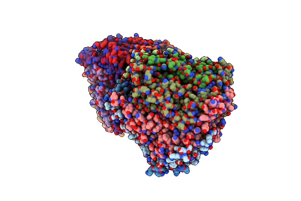
Organism: Plasmodium falciparum (isolate 3d7), Plasmodium falciparum
Method: ELECTRON MICROSCOPY Release Date: 2021-09-22 Classification: HYDROLASE Ligands: BO2 |
 |
Organism: Plasmodium falciparum (isolate 3d7)
Method: ELECTRON MICROSCOPY Release Date: 2021-09-22 Classification: HYDROLASE Ligands: YHD |
 |
Organism: Homo sapiens
Method: ELECTRON MICROSCOPY Release Date: 2021-09-22 Classification: HYDROLASE Ligands: YHD |
 |
Organism: Mycolicibacterium smegmatis (strain atcc 700084 / mc(2)155)
Method: X-RAY DIFFRACTION Resolution:2.30 Å Release Date: 2020-05-13 Classification: BIOSYNTHETIC PROTEIN Ligands: CA |
 |
Organism: Mycolicibacterium smegmatis (strain atcc 700084 / mc(2)155)
Method: X-RAY DIFFRACTION Resolution:2.21 Å Release Date: 2020-05-13 Classification: BIOSYNTHETIC PROTEIN Ligands: FO1, CA |
 |
Organism: Mycolicibacterium smegmatis (strain atcc 700084 / mc(2)155)
Method: X-RAY DIFFRACTION Resolution:2.40 Å Release Date: 2020-05-13 Classification: BIOSYNTHETIC PROTEIN Ligands: GDP, GOL, CA |
 |
The Crystal Structure Of Fbia From Mycobacterium Smegmatis, Gdp And Fo Bound Form
Organism: Mycolicibacterium smegmatis (strain atcc 700084 / mc(2)155)
Method: X-RAY DIFFRACTION Resolution:2.20 Å Release Date: 2020-05-13 Classification: BIOSYNTHETIC PROTEIN Ligands: GDP, FO1, CA |
 |
The Crystal Structure Of Fbia From Mycobacterium Smegmatis, Dehydro-F420-0 Bound Form
Organism: Mycolicibacterium smegmatis (strain atcc 700084 / mc(2)155)
Method: X-RAY DIFFRACTION Resolution:2.34 Å Release Date: 2020-05-13 Classification: BIOSYNTHETIC PROTEIN Ligands: GOL, QMD, CA |
 |
Crystal Structure Of The 11S Subunit Of The Plasmodium Falciparum Proteasome, Pa28
Organism: Plasmodium falciparum
Method: X-RAY DIFFRACTION Resolution:3.10 Å Release Date: 2019-08-07 Classification: PROTEIN BINDING Ligands: SO4 |
 |
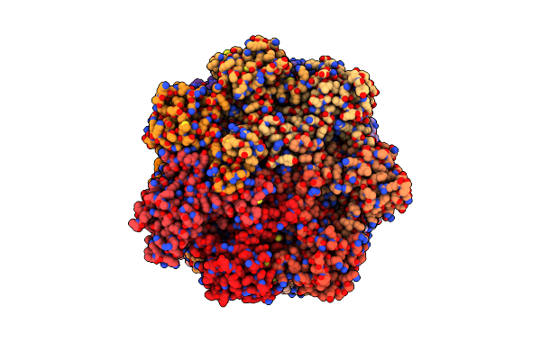
The Structure Of The Plasmodium Falciparum 20S Proteasome In Complex With Two Pa28 Activators
Organism: Plasmodium falciparum (isolate 3d7)
Method: ELECTRON MICROSCOPY Release Date: 2019-08-07 Classification: HYDROLASE |
 |
Organism: Plasmodium falciparum (isolate 3d7)
Method: ELECTRON MICROSCOPY Release Date: 2019-08-07 Classification: HYDROLASE |
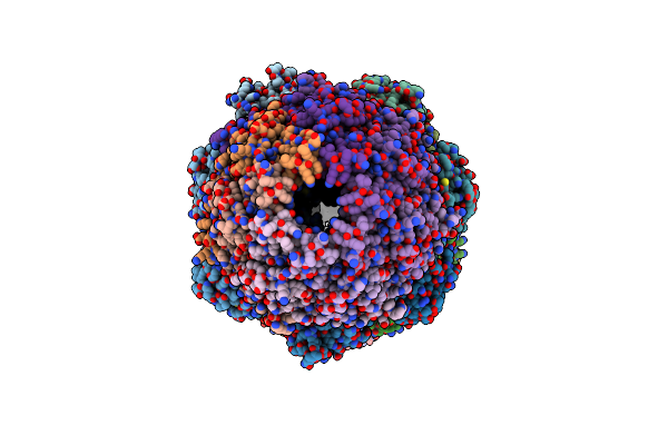 |
The Structure Of The Plasmodium Falciparum 20S Proteasome In Complex With One Pa28 Activator
Organism: Plasmodium falciparum 3d7
Method: ELECTRON MICROSCOPY Release Date: 2019-08-07 Classification: HYDROLASE |