Planned Maintenance: Some services may turn out to be unavailable from 15th January, 2026 to 16th January, 2026. We apologize for the inconvenience!

Planned Maintenance: Some services may turn out to be unavailable from 15th January, 2026 to 16th January, 2026. We apologize for the inconvenience!
 |
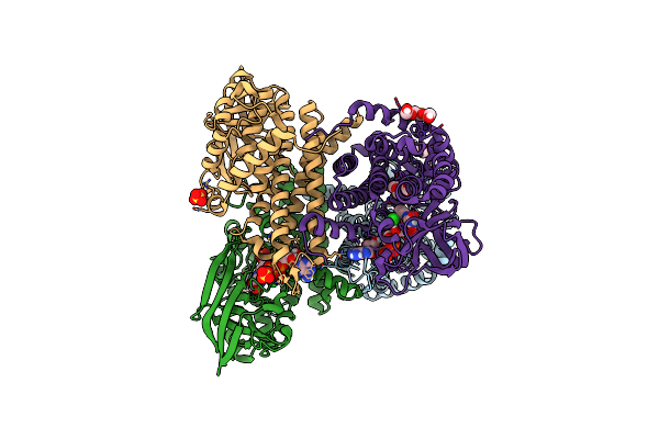
Structure Of The Pel Modification Enzyme Pela From Pseudomonas Thermotolerans
Organism: Pseudomonas thermotolerans dsm 14292
Method: X-RAY DIFFRACTION Resolution:2.09 Å Release Date: 2025-05-14 Classification: HYDROLASE |
 |
Crystal Structure Of Mycobacterium Tuberculosis Cytochrome P450 Cyp125 In Complex With An Inhibitor
Organism: Mycobacterium tuberculosis h37rv
Method: X-RAY DIFFRACTION Release Date: 2025-01-29 Classification: OXIDOREDUCTASE Ligands: HEM, SO4, A1H47 |
 |
X-Ray Crystal Structure Of Cyp142 From Mycobacterium Tuberculosis In Complex With A Fragment Bound In Two Poses
Organism: Mycobacterium tuberculosis h37rv
Method: X-RAY DIFFRACTION Release Date: 2025-01-29 Classification: OXIDOREDUCTASE Ligands: 3QO, HEM |
 |
Crystal Structure Of Cyp125 From Mycobacterium Tuberculosis In Complex With An Inhibitor
Organism: Mycobacterium tuberculosis h37rv
Method: X-RAY DIFFRACTION Release Date: 2023-04-19 Classification: OXIDOREDUCTASE Ligands: EIQ, HEM, CL, DMS |
 |
Crystal Structure Of Cyp125 From Mycobacterium Tuberculosis In Complex With An Inhibitor
Organism: Mycobacterium tuberculosis h37rv
Method: X-RAY DIFFRACTION Release Date: 2023-04-19 Classification: OXIDOREDUCTASE Ligands: GOL, 5YG, HEM, SO4, CL |
 |
Crystal Structure Of Cyp142 From Mycobacterium Tuberculosis In Complex With An Inhibitor At Partial Occupancy With Peg
Organism: Mycobacterium tuberculosis h37rv
Method: X-RAY DIFFRACTION Release Date: 2023-01-18 Classification: OXIDOREDUCTASE Ligands: HEM, ACT, PG4, BR, CL, EIQ |
 |
Structure Of Cyp142 From Mycobacterium Tuberculosis In Complex With Inhibitor Mek216
Organism: Mycobacterium tuberculosis h37rv
Method: X-RAY DIFFRACTION Release Date: 2022-11-16 Classification: OXIDOREDUCTASE Ligands: HEM, 5YG, BR, K |
 |
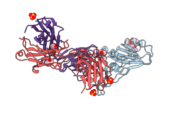
Crystal Structure Of The Receptor Binding Domain Of Sars-Cov-2 Spike Glycoprotein In Complex With Covox-222 And Covox-278 Fabs
Organism: Severe acute respiratory syndrome coronavirus 2, Homo sapiens
Method: X-RAY DIFFRACTION Resolution:2.34 Å Release Date: 2021-07-07 Classification: VIRAL PROTEIN Ligands: GOL, CL, PG4 |
 |
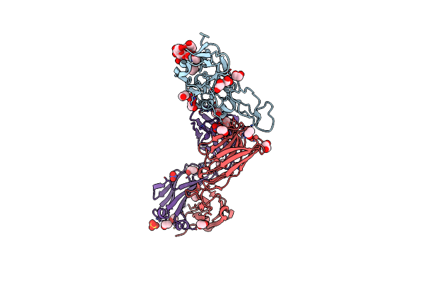
Crystal Structure Of The T478K Mutant Receptor Binding Domain Of Sars-Cov-2 Spike Glycoprotein In Complex With Covox-45 And Covox-253 Fabs
Organism: Severe acute respiratory syndrome coronavirus 2, Homo sapiens
Method: X-RAY DIFFRACTION Resolution:2.60 Å Release Date: 2021-07-07 Classification: VIRAL PROTEIN Ligands: GOL, CL |
 |
Crystal Structure Of The L452R Mutant Receptor Binding Domain Of Sars-Cov-2 Spike Glycoprotein In Complex With Covox-75 And Covox-253 Fabs
Organism: Severe acute respiratory syndrome coronavirus 2, Homo sapiens
Method: X-RAY DIFFRACTION Resolution:2.50 Å Release Date: 2021-07-07 Classification: VIRAL PROTEIN Ligands: GOL, PEG, CL, TRS, PO4, BTB |
 |
Crystal Structure Of The Receptor Binding Domain Of Sars-Cov-2 Spike Glycoprotein In Complex With Covox-222 And Ey6A Fabs
Organism: Homo sapiens, Severe acute respiratory syndrome coronavirus 2
Method: X-RAY DIFFRACTION Resolution:2.25 Å Release Date: 2021-04-07 Classification: VIRAL PROTEIN/IMMUNE SYSTEM Ligands: CL, SO4, GOL, NAG |
 |
Crystal Structure Of The K417N Mutant Receptor Binding Domain Of Sars-Cov-2 Spike Glycoprotein In Complex With Covox-222 And Ey6A Fabs
Organism: Homo sapiens, Severe acute respiratory syndrome coronavirus 2
Method: X-RAY DIFFRACTION Resolution:2.30 Å Release Date: 2021-04-07 Classification: VIRAL PROTEIN/IMMUNE SYSTEM Ligands: GOL, NAG, SO4, CL, CIT |
 |
Crystal Structure Of The K417T Mutant Receptor Binding Domain Of Sars-Cov-2 Spike Glycoprotein In Complex With Covox-222 And Ey6A Fabs
Organism: Homo sapiens, Severe acute respiratory syndrome coronavirus 2
Method: X-RAY DIFFRACTION Resolution:1.95 Å Release Date: 2021-04-07 Classification: VIRAL PROTEIN/IMMUNE SYSTEM Ligands: NAG, GOL, PEG, SO4, CIT, CL |
 |
Crystal Structure Of The N501Y Mutant Receptor Binding Domain Of Sars-Cov-2 Spike Glycoprotein In Complex With Covox-222 And Ey6A Fabs
Organism: Homo sapiens, Severe acute respiratory syndrome coronavirus 2
Method: X-RAY DIFFRACTION Resolution:2.40 Å Release Date: 2021-04-07 Classification: VIRAL PROTEIN/IMMUNE SYSTEM Ligands: SO4, GOL, NAG, PEG, CL |
 |
Crystal Structure Of The Receptor Binding Domain Of Sars-Cov-2 B.1.351 Variant Spike Glycoprotein In Complex With Covox-222 And Ey6A Fabs
Organism: Homo sapiens, Severe acute respiratory syndrome coronavirus 2
Method: X-RAY DIFFRACTION Resolution:2.50 Å Release Date: 2021-04-07 Classification: VIRAL PROTEIN/IMMUNE SYSTEM Ligands: SO4, GOL, NAG, PEG |
 |
Crystal Structure Of The Receptor Binding Domain Of Sars-Cov-2 P.1 Variant Spike Glycoprotein In Complex With Covox-222 And Ey6A Fabs
Organism: Homo sapiens, Severe acute respiratory syndrome coronavirus 2
Method: X-RAY DIFFRACTION Resolution:2.67 Å Release Date: 2021-04-07 Classification: VIRAL PROTEIN/IMMUNE SYSTEM Ligands: SO4, GOL, NAG, PEG |
 |
Crystal Structure Of The Receptor Binding Domain Of Sars-Cov-2 P.1 Variant Spike Glycoprotein In Complex With Ace2
Organism: Homo sapiens, Severe acute respiratory syndrome coronavirus 2
Method: X-RAY DIFFRACTION Resolution:3.14 Å Release Date: 2021-04-07 Classification: VIRAL PROTEIN/HYDROLASE Ligands: NAG |
 |
Organism: Thermomonospora curvata (strain atcc 19995 / dsm 43183 / jcm 3096 / nbrc 15933 / ncimb 10081 / henssen b9)
Method: X-RAY DIFFRACTION Resolution:2.10 Å Release Date: 2021-03-24 Classification: OXIDOREDUCTASE Ligands: PGE, FAD, CL, MES, SO4 |
 |
Crystal Structure Of The N501Y Mutant Receptor Binding Domain Of Sars-Cov-2 Spike Glycoprotein In Complex With Covox-269 Fab
Organism: Homo sapiens, Severe acute respiratory syndrome coronavirus 2
Method: X-RAY DIFFRACTION Resolution:2.19 Å Release Date: 2021-03-03 Classification: VIRAL PROTEIN/IMMUNE SYSTEM Ligands: GOL, SO4 |
 |
Crystal Structure Of The Receptor Binding Domain Of Sars-Cov-2 Spike Glycoprotein In Complex With Covox-269 Fab
Organism: Homo sapiens, Severe acute respiratory syndrome coronavirus 2
Method: X-RAY DIFFRACTION Release Date: 2021-03-03 Classification: VIRAL PROTEIN/IMMUNE SYSTEM Ligands: EDO, NO3, PEG, SO4, CL |