
Search Count: 21
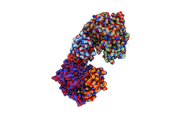 |
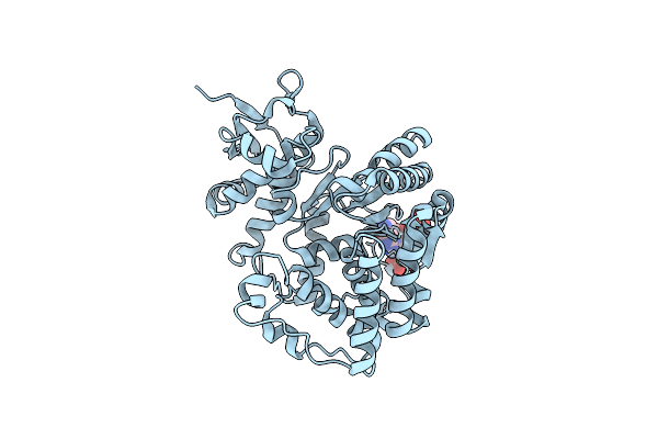
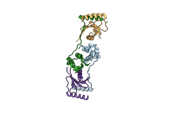
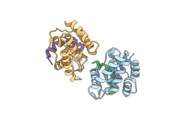
Crystal Structure Of The Escherichia Coli Toxin-Antitoxin System Hipbst (Hipt S57A)
Organism: Escherichia coli o127:h6 (strain e2348/69 / epec)
Method: X-RAY DIFFRACTION Resolution:2.40 Å Release Date: 2022-01-12 Classification: TOXIN |
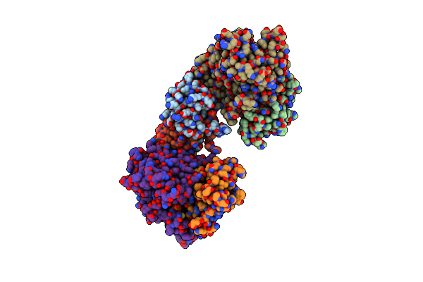 |
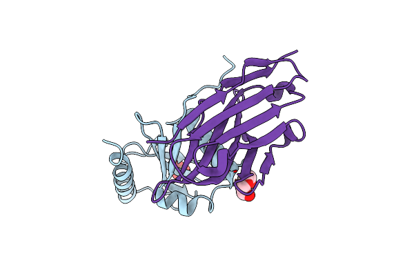
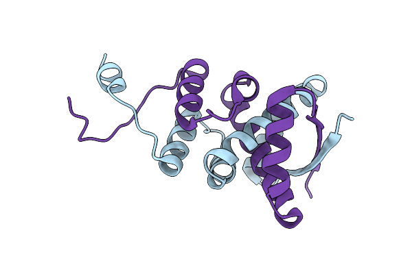
Crystal Structure Of The Escherichia Coli Toxin-Antitoxin System Hipbst (Hipt S59A)
Organism: Escherichia coli o127:h6 (strain e2348/69 / epec)
Method: X-RAY DIFFRACTION Resolution:3.34 Å Release Date: 2022-01-12 Classification: TOXIN |
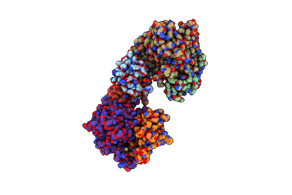 |
Crystal Structure Of The Escherichia Coli Toxin-Antitoxin System Hipbst (Hipt D233Q)
Organism: Escherichia coli o127:h6 (strain e2348/69 / epec)
Method: X-RAY DIFFRACTION Resolution:2.90 Å Release Date: 2022-01-12 Classification: TOXIN |
 |
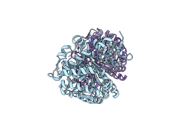
Organism: Escherichia coli str. k-12 substr. mg1655, Escherichia coli 2-222-05_s4_c3
Method: X-RAY DIFFRACTION Resolution:2.28 Å Release Date: 2019-09-18 Classification: TOXIN Ligands: SO4 |
 |
Organism: Escherichia coli 2-210-07_s3_c3
Method: X-RAY DIFFRACTION Resolution:2.26 Å Release Date: 2019-09-18 Classification: ANTITOXIN |
 |
Organism: Escherichia coli k-12
Method: X-RAY DIFFRACTION Resolution:2.48 Å Release Date: 2019-04-24 Classification: HYDROLASE |
 |
Organism: Escherichia coli k-12
Method: X-RAY DIFFRACTION Resolution:2.77 Å Release Date: 2019-04-24 Classification: HYDROLASE Ligands: 0O2 |
 |
Organism: Pseudomonas putida kt2440
Method: X-RAY DIFFRACTION Resolution:2.21 Å Release Date: 2018-10-24 Classification: TOXIN Ligands: IMD, GOL |
 |
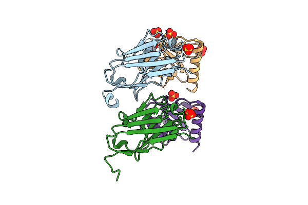
Organism: Vibrio cholerae
Method: X-RAY DIFFRACTION Resolution:1.80 Å Release Date: 2017-04-05 Classification: ANTITOXIN |
 |
Organism: Vibrio cholerae serotype o1 (strain atcc 39315 / el tor inaba n16961), Lama glama
Method: X-RAY DIFFRACTION Resolution:2.49 Å Release Date: 2017-04-05 Classification: TOXIN Ligands: PO4, ACT, EDO, EPE, PDO |
 |
Organism: Vibrio cholerae serotype o1 (strain atcc 39315 / el tor inaba n16961), Lama glama
Method: X-RAY DIFFRACTION Resolution:1.85 Å Release Date: 2017-04-05 Classification: TOXIN Ligands: SO4, EDO |
 |
Organism: Vibrio cholerae serotype o1 (strain atcc 39315 / el tor inaba n16961)
Method: X-RAY DIFFRACTION Resolution:2.99 Å Release Date: 2017-04-05 Classification: TOXIN |
 |
Organism: Vibrio cholerae, Lama glama
Method: X-RAY DIFFRACTION Release Date: 2017-04-05 Classification: TOXIN Ligands: PEG, SO4 |
 |
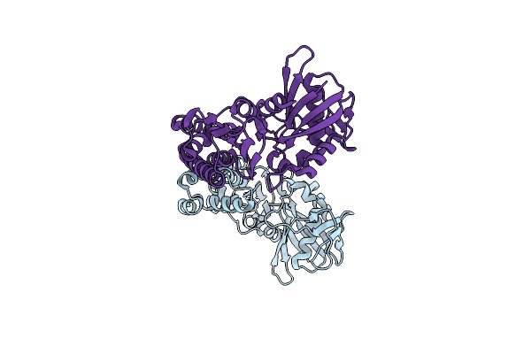
Organism: Escherichia coli k-12, Thermus thermophilus hb8, Thermus thermophilus
Method: X-RAY DIFFRACTION Resolution:3.30 Å Release Date: 2014-07-09 Classification: RIBOSOME Ligands: ZN, MG |
 |
Organism: Escherichia coli k-12, Thermus thermophilus hb8, Thermus thermophilus
Method: X-RAY DIFFRACTION Resolution:3.60 Å Release Date: 2014-07-09 Classification: RIBOSOME Ligands: ZN, MG |
 |
Organism: Shigella flexneri
Method: X-RAY DIFFRACTION Resolution:2.70 Å Release Date: 2011-11-02 Classification: TRANSLATION, TOXIN Ligands: SO4, NA |
 |
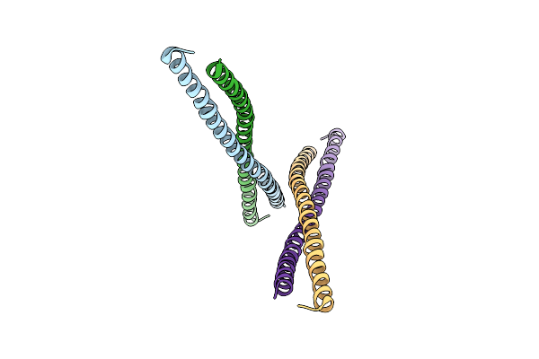
Organism: Enterobacteria phage p1
Method: X-RAY DIFFRACTION Resolution:1.70 Å Release Date: 2008-09-16 Classification: RIBOSOME INHIBITOR Ligands: BR |
 |
Organism: Escherichia coli
Method: X-RAY DIFFRACTION Resolution:2.80 Å Release Date: 2008-02-05 Classification: CELL CYCLE |
 |
Organism: Escherichia coli
Method: X-RAY DIFFRACTION Resolution:2.80 Å Release Date: 2007-10-09 Classification: DNA BINDING PROTEIN |
 |
Organism: Escherichia coli
Method: X-RAY DIFFRACTION Resolution:2.30 Å Release Date: 2003-01-28 Classification: STRUCTURAL PROTEIN |