
Search Count: 18
 |
Crystal Structure Of The Core Fragment Of Muty From E.Coli At 1.2A Resolution
Organism: Escherichia coli
Method: X-RAY DIFFRACTION Resolution:1.20 Å Release Date: 2002-11-26 Classification: HYDROLASE Ligands: SO4, SF4, GOL |
 |
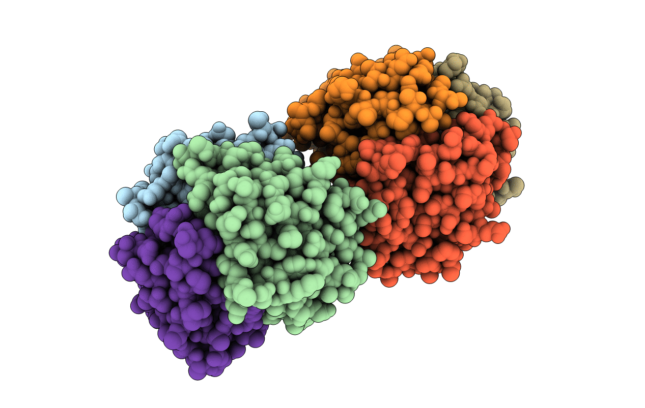
Crystal Structure Of The Core Fragment Of Muty From E.Coli At 1.55A Resolution
Organism: Escherichia coli
Method: X-RAY DIFFRACTION Resolution:1.55 Å Release Date: 2002-11-26 Classification: HYDROLASE Ligands: SO4, SF4, GOL |
 |
Organism: Escherichia coli
Method: X-RAY DIFFRACTION Resolution:1.60 Å Release Date: 2002-11-26 Classification: HYDROLASE Ligands: SO4, SF4, GOL |
 |
Organism: Escherichia coli
Method: X-RAY DIFFRACTION Resolution:1.35 Å Release Date: 2002-11-26 Classification: HYDROLASE Ligands: SO4, SF4, GOL |
 |
Organism: Escherichia coli
Method: X-RAY DIFFRACTION Resolution:1.50 Å Release Date: 2002-11-26 Classification: HYDROLASE Ligands: SO4, SF4, GOL |
 |
Organism: Escherichia coli
Method: X-RAY DIFFRACTION Resolution:1.50 Å Release Date: 2002-11-26 Classification: HYDROLASE Ligands: SO4, SF4, GOL |
 |
Crystal Structure Of A Trapped Reaction Intermediate Of The Dna Repair Enzyme Endonuclease Viii With Dna
Organism: Escherichia coli
Method: X-RAY DIFFRACTION Resolution:1.42 Å Release Date: 2002-10-04 Classification: HYDROLASE/DNA Ligands: ZN, SO4 |
 |
Crystal Structure Of A Trapped Reaction Intermediate Of The Dna Repair Enzyme Endonuclease Viii With Brominated-Dna
Organism: Escherichia coli
Method: X-RAY DIFFRACTION Resolution:1.25 Å Release Date: 2002-10-04 Classification: HYDROLASE/DNA Ligands: ZN, SO4, GOL |
 |
Crystal Structure Of E.Coli Formamidopyrimidine-Dna Glycosylase (Fpg) Covalently Trapped With Dna
Organism: Escherichia coli
Method: X-RAY DIFFRACTION Resolution:2.10 Å Release Date: 2002-06-14 Classification: HYDROLASE/DNA Ligands: ZN |
 |
Crystal Structure Of Saccharomyces Cerevisiae Nit3, A Member Of Branch 10 Of The Nitrilase Superfamily
Organism: Saccharomyces cerevisiae
Method: X-RAY DIFFRACTION Resolution:2.40 Å Release Date: 2001-10-04 Classification: STRUCTURAL GENOMICS, UNKNOWN FUNCTION |
 |
Organism: Saccharomyces cerevisiae
Method: X-RAY DIFFRACTION Resolution:1.94 Å Release Date: 2001-09-26 Classification: structural genomics, unknown function Ligands: CL |
 |
Organism: Saccharomyces cerevisiae
Method: X-RAY DIFFRACTION Resolution:1.70 Å Release Date: 2001-07-04 Classification: TRANSLATION INHIBITOR |
 |
Structural Studies Of Cholesterol Biosynthesis: Mevalonate 5-Diphosphate Decarboxylase And Isopentenyl Diphosphate Isomerase
Organism: Escherichia coli
Method: X-RAY DIFFRACTION Resolution:2.50 Å Release Date: 2001-03-28 Classification: ISOMERASE Ligands: MN |
 |
The X-Ray Crystal Structure Of Mevalonate 5-Diphosphate Decarboxylase At 2.3 Angstrom Resolution.
Organism: Saccharomyces cerevisiae
Method: X-RAY DIFFRACTION Resolution:2.27 Å Release Date: 2001-03-21 Classification: LYASE |
 |
Organism: Saccharomyces cerevisiae
Method: X-RAY DIFFRACTION Resolution:2.00 Å Release Date: 1999-09-02 Classification: STRUCTURAL GENOMICS Ligands: PLP |
 |
Organism: Saccharomyces cerevisiae
Method: X-RAY DIFFRACTION Resolution:2.70 Å Release Date: 1999-08-25 Classification: OXIDOREDUCTASE Ligands: FMN |
 |
Crystal Structure Of A Yeast Hypothetical Protein-A Structure From Bnl'S Human Proteome Project
Organism: Saccharomyces cerevisiae
Method: X-RAY DIFFRACTION Resolution:2.10 Å Release Date: 1999-01-27 Classification: HYPOTHETICAL PROTEIN Ligands: PLP |
 |
Organism: Geobacillus stearothermophilus
Method: X-RAY DIFFRACTION Resolution:2.60 Å Release Date: 1997-01-11 Classification: RIBOSOMAL PROTEIN |