
Search Count: 28
 |
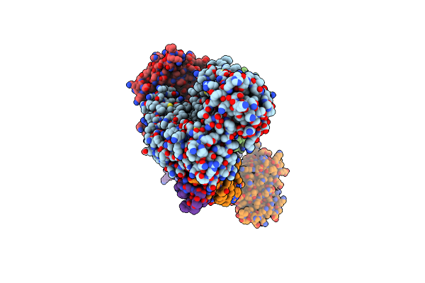
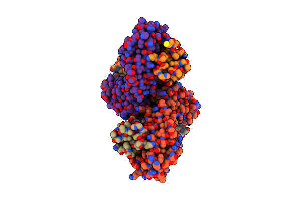
Organism: Homo sapiens, Lama glama
Method: X-RAY DIFFRACTION Release Date: 2025-10-29 Classification: PROTEIN TRANSPORT |
 |
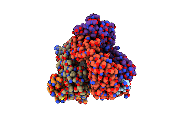
Organism: Bacillus subtilis subsp. subtilis str. 168
Method: ELECTRON MICROSCOPY Release Date: 2023-06-21 Classification: MEMBRANE PROTEIN Ligands: PLM, 6OU, OLA |
 |
Organism: Bacillus subtilis subsp. subtilis str. 168
Method: ELECTRON MICROSCOPY Release Date: 2023-06-21 Classification: MEMBRANE PROTEIN Ligands: OLA, 6OU |
 |
Organism: Bacillus subtilis subsp. subtilis str. 168
Method: ELECTRON MICROSCOPY Release Date: 2023-06-21 Classification: MEMBRANE PROTEIN Ligands: PLM, 6OU, OLA |
 |
Organism: Bacillus subtilis subsp. subtilis str. 168
Method: ELECTRON MICROSCOPY Release Date: 2023-06-21 Classification: MEMBRANE PROTEIN Ligands: PLM, 6OU, OLA |
 |
Organism: Bacillus subtilis subsp. subtilis str. 168
Method: ELECTRON MICROSCOPY Release Date: 2023-06-21 Classification: MEMBRANE PROTEIN Ligands: 6OU, AGS |
 |
Organism: Bacillus subtilis subsp. subtilis str. 168
Method: ELECTRON MICROSCOPY Release Date: 2023-06-21 Classification: MEMBRANE PROTEIN Ligands: AGS |
 |
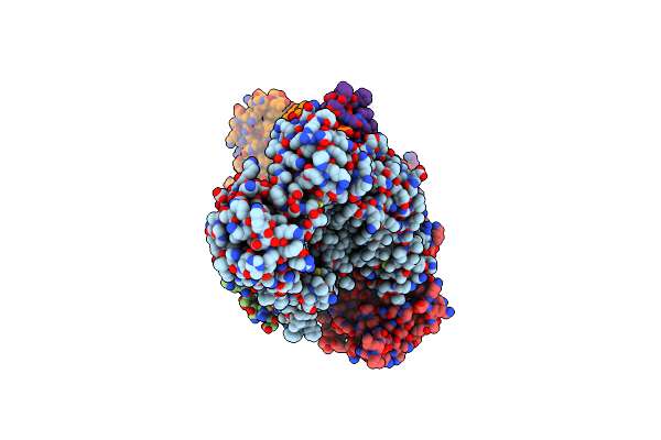
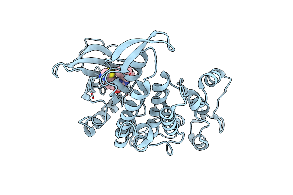
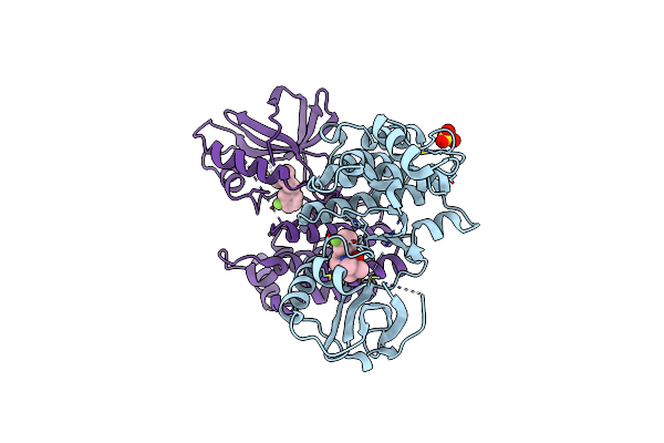
Organism: Homo sapiens
Method: X-RAY DIFFRACTION Resolution:2.60 Å Release Date: 2023-05-17 Classification: TRANSFERASE Ligands: WAK |
 |
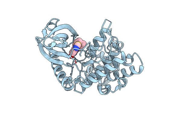
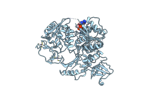
Crystal Structure Of Human Casein Kinase Ii Subunit Alpha (Ck2A1) In Complex With Leucettinib-92
Organism: Homo sapiens
Method: X-RAY DIFFRACTION Resolution:2.45 Å Release Date: 2023-05-17 Classification: TRANSFERASE Ligands: SO4, WAK |
 |
Organism: Homo sapiens
Method: X-RAY DIFFRACTION Resolution:2.40 Å Release Date: 2023-05-17 Classification: TRANSFERASE Ligands: WAZ |
 |
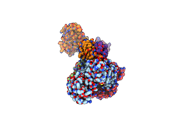
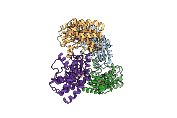
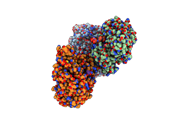
Organism: Bacillus subtilis subsp. subtilis str. 168
Method: ELECTRON MICROSCOPY Release Date: 2022-04-06 Classification: TRANSPORT PROTEIN Ligands: I0O |
 |
Organism: Bacillus subtilis subsp. subtilis str. 168
Method: ELECTRON MICROSCOPY Release Date: 2022-04-06 Classification: TRANSPORT PROTEIN Ligands: I0O, ATP |
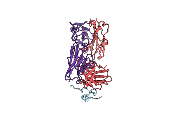 |
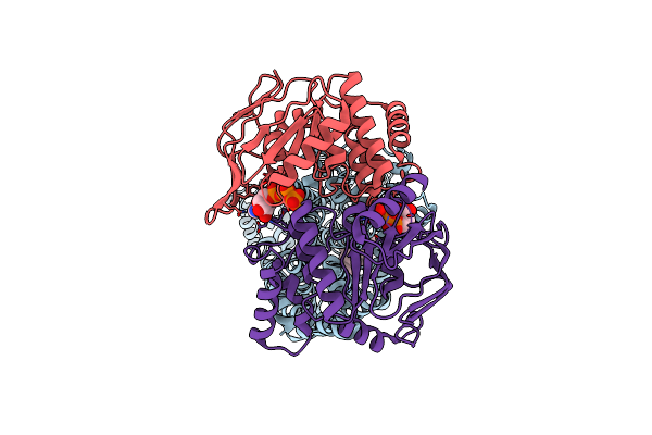
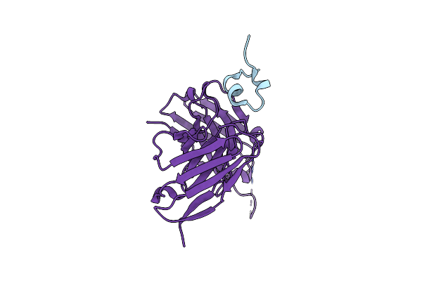
Organism: Homo sapiens, Human respiratory syncytial virus a (strain a2)
Method: X-RAY DIFFRACTION Resolution:2.90 Å Release Date: 2019-12-18 Classification: VIRAL PROTEIN, IMMUNE SYSTEM |
 |
Organism: Homo sapiens
Method: X-RAY DIFFRACTION Resolution:2.79 Å Release Date: 2019-05-08 Classification: SIGNALING PROTEIN/INHIBITOR Ligands: M5J, SO4 |
 |
Organism: Homo sapiens
Method: X-RAY DIFFRACTION Resolution:3.25 Å Release Date: 2019-05-01 Classification: TRANSFERASE Ligands: JSW |
 |
Organism: Homo sapiens, Human respiratory syncytial virus a (strain a2)
Method: X-RAY DIFFRACTION Resolution:1.55 Å Release Date: 2018-03-14 Classification: VIRAL PROTEIN/IMMUNE SYSTEM |
 |
Organism: Homo sapiens, Human respiratory syncytial virus a
Method: X-RAY DIFFRACTION Resolution:2.40 Å Release Date: 2018-03-14 Classification: VIRAL PROTEIN/IMMUNE SYSTEM |
 |
Organism: Homo sapiens, Human respiratory syncytial virus a
Method: X-RAY DIFFRACTION Resolution:2.40 Å Release Date: 2018-03-14 Classification: VIRAL PROTEIN/IMMUNE SYSTEM Ligands: ZN, ACY |
 |
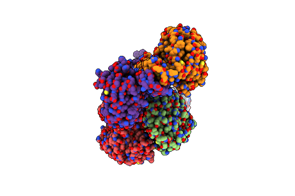
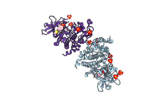
Organism: Klebsiella pneumoniae subsp. pneumoniae
Method: X-RAY DIFFRACTION Resolution:2.65 Å Release Date: 2014-01-08 Classification: DNA BINDING PROTEIN Ligands: ADP, ZN |
 |
Organism: Klebsiella pneumoniae subsp. pneumoniae
Method: X-RAY DIFFRACTION Resolution:4.08 Å Release Date: 2014-01-08 Classification: DNA BINDING PROTEIN Ligands: ZN |