
Search Count: 25
 |
Organism: Severe acute respiratory syndrome coronavirus 2
Method: X-RAY DIFFRACTION Resolution:1.90 Å Release Date: 2025-04-23 Classification: VIRAL PROTEIN Ligands: A1IYL |
 |
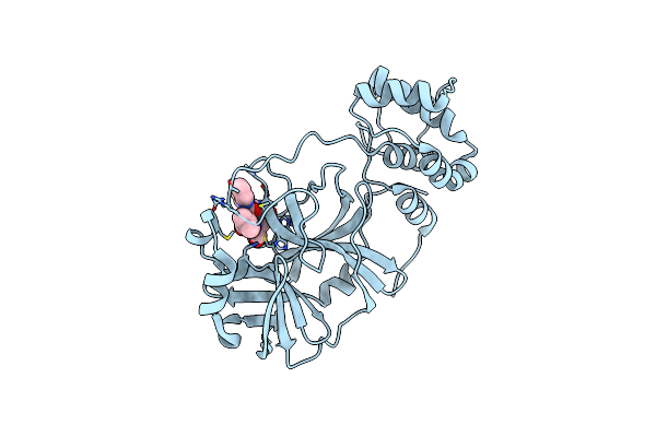
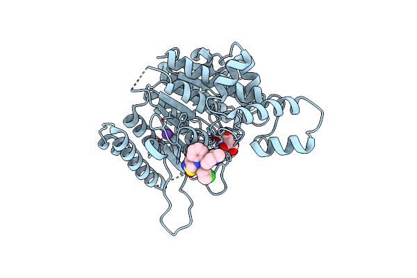
Complex Crystal Structure Of Mutant Human Monoglyceride Lipase With Compound 5D
Organism: Homo sapiens
Method: X-RAY DIFFRACTION Resolution:1.51 Å Release Date: 2024-01-31 Classification: HYDROLASE Ligands: F28, EDO |
 |
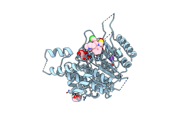
Complex Crystal Structure Of Mutant Human Monoglyceride Lipase With Compound 5L
Organism: Homo sapiens
Method: X-RAY DIFFRACTION Resolution:1.55 Å Release Date: 2024-01-31 Classification: HYDROLASE Ligands: EJI, EDO |
 |
Complex Crystal Structure Of Mutant Human Monoglyceride Lipase With Compound 5R
Organism: Homo sapiens
Method: X-RAY DIFFRACTION Resolution:1.73 Å Release Date: 2024-01-31 Classification: HYDROLASE Ligands: EFH, EDO |
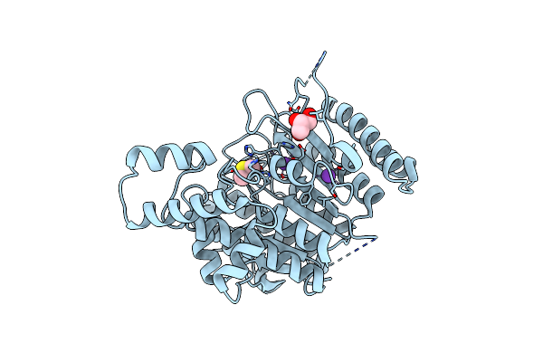 |
Crystal Structure Of Schistosoma Mansoni Hdac8 With Dmso Bound In The Active Site
Organism: Schistosoma mansoni
Method: X-RAY DIFFRACTION Resolution:1.90 Å Release Date: 2022-08-31 Classification: HYDROLASE Ligands: ZN, K, GOL, DMS |
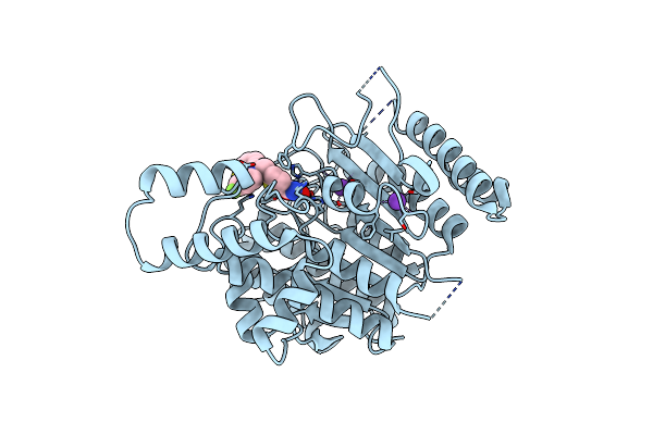 |
Crystal Structure Of Schistosoma Mansoni Hdac8 In Complex With A Tricyclic Thieno[3,2-B]Indole Capped Hydroxamate-Based Inhibitor, Chlorine Derivative
Organism: Schistosoma mansoni
Method: X-RAY DIFFRACTION Resolution:2.20 Å Release Date: 2022-07-27 Classification: HYDROLASE Ligands: ZN, K, 4UI |
 |
Tetartohedrally Twinned Crystal Structure Of Schistosoma Mansoni Hdac8 In Complex With A Tricyclic Thieno[3,2-B]Indole Capped Hydroxamate-Based Inhibitor, Bromine Derivative
Organism: Schistosoma mansoni
Method: X-RAY DIFFRACTION Resolution:2.30 Å Release Date: 2022-07-20 Classification: HYDROLASE Ligands: ZN, K, 4VX, 144, CL |
 |
Crystal Structure Of Schistosoma Mansoni Hdac8 In Complex With A 3-Chlorophenyl-Spiroindoline Capped Hydroxamate-Based Inhibitor, Bound To A Novel Site
Organism: Schistosoma mansoni
Method: X-RAY DIFFRACTION Resolution:1.80 Å Release Date: 2022-07-20 Classification: HYDROLASE Ligands: ZN, K, CL, TLA, 4WB |
 |
Crystal Structure Of Schistosoma Mansoni Hdac8 In Complex With A 4-Chlorophenyl-Spiroindoline Capped Hydroxamate-Based Inhibitor, Bound To A Novel Site
Organism: Schistosoma mansoni
Method: X-RAY DIFFRACTION Resolution:2.25 Å Release Date: 2022-07-20 Classification: HYDROLASE Ligands: ZN, K, TLA, 4XI, PEG |
 |
Trypanothione Reductase From Trypanosoma Brucei In Complex With N-{4-Methoxy-3-[(4-Methoxyphenyl)Sulfamoyl]Phenyl}-5-Nitrothiophene-2-Carboxamide
Organism: Trypanosoma brucei brucei (strain 927/4 gutat10.1)
Method: X-RAY DIFFRACTION Resolution:2.15 Å Release Date: 2022-03-30 Classification: OXIDOREDUCTASE Ligands: FAD, UT2, GOL, SO4 |
 |
Crystal Structure Of Danio Rerio Histone Deacetylase 6 Catalytic Domain 2 (Cd2) Complexed With Nf2376
Organism: Danio rerio
Method: X-RAY DIFFRACTION Resolution:2.04 Å Release Date: 2020-12-02 Classification: HYDROLASE Ligands: ZN, K, QQD, EDO |
 |
Crystal Structure Of Danio Rerio Histone Deacetylase 6 Catalytic Domain 2 (Cd2) Complexed With Nf2657
Organism: Danio rerio
Method: X-RAY DIFFRACTION Resolution:2.09 Å Release Date: 2020-12-02 Classification: HYDROLASE Ligands: ZN, K, QQG |
 |
Structure Of The Ampa Receptor Glua2O Ligand-Binding Domain (S1S2J) In Complex With The Compound (S)-1-[2'-Amino-2'-Carboxyethyl]-5,7-Dihydrothieno[3,4-D]Pyrimidin- 2,4(1H,3H)-Dione At Resolution 1.60A
Organism: Rattus norvegicus
Method: X-RAY DIFFRACTION Resolution:1.61 Å Release Date: 2020-06-03 Classification: MEMBRANE PROTEIN Ligands: SO4, GOL, CGW, CL |
 |
Structure Of The Ampa Receptor Glua2O Ligand-Binding Domain (S1S2J) In Complex With The Compound ( S) - 1- [2'-Amino-2'-Carboxyethyl]-5 ,7- Dihydropyrrolo[3,4-D]Pyrimidin-2,4(1H,3H)-Dione At Resolution 1.20A
Organism: Rattus norvegicus
Method: X-RAY DIFFRACTION Resolution:1.20 Å Release Date: 2020-06-03 Classification: MEMBRANE PROTEIN Ligands: GOL, SO4, PVQ, NH4, CL |
 |
Structure Of The Ampa Receptor Glua2O Ligand-Binding Domain (S1S2J) In Complex With The Compound ( S) - 1- [2'-Amino-2'-Carboxyethyl]-6-Methyl-5 ,7- Dihydropyrrolo[3,4-D]Pyrimidin-2,4(1H,3H)-Dione At Resolution 1.00A
Organism: Rattus norvegicus
Method: X-RAY DIFFRACTION Resolution:1.00 Å Release Date: 2020-06-03 Classification: MEMBRANE PROTEIN Ligands: SO4, GOL, LI, CG8, CL |
 |
Structure Of The Ampa Receptor Glua2O Ligand-Binding Domain (S1S2J) In Complex With The Compound (S)-1-(2'-Amino-2'-Carboxyethyl)-5,7-Dihydrofuro[3,4-D]- Pyrimidine-2,4(1H,3H)-Dione At Resolution 1.15A
Organism: Rattus norvegicus
Method: X-RAY DIFFRACTION Resolution:1.15 Å Release Date: 2020-06-03 Classification: MEMBRANE PROTEIN Ligands: GOL, SO4, LI, PVK, CL |
 |
Structure Of The Ampa Receptor Glua2O Ligand-Binding Domain (S1S2J) In Complex With The Compound (S)-1-(2'-Amino-2'-Carboxyethyl)Furo[3,4-D]Pyrimidin-2,4-Dione At Resolution 1.47A
Organism: Rattus norvegicus
Method: X-RAY DIFFRACTION Resolution:1.47 Å Release Date: 2020-06-03 Classification: MEMBRANE PROTEIN Ligands: SO4, GOL, LI, CL, OUB |
 |
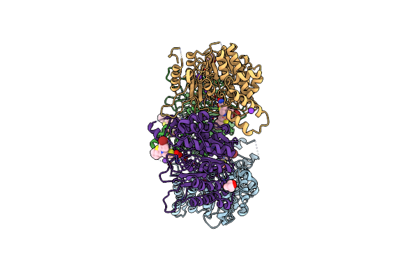
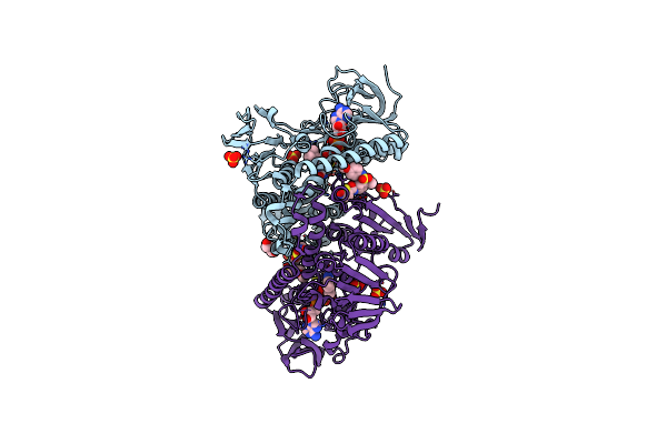
Organism: Rattus norvegicus, Gallus gallus, Bos taurus
Method: X-RAY DIFFRACTION Resolution:2.40 Å Release Date: 2018-12-05 Classification: CELL CYCLE Ligands: GTP, MG, CA, GDP, EZW, MES, ACP |
 |
Crystal Structure Of The Kainate Receptor Gluk3 Ligand Binding Domain In Complex With (S)-1-[2'-Amino-2'-Carboxyethyl]-6-Methyl-5,7-Dihydropyrrolo[3,4-D]Pyrimidin-2,4(1H,3H)-Dione At Resolution 2.4A
Organism: Rattus norvegicus
Method: X-RAY DIFFRACTION Resolution:2.40 Å Release Date: 2018-02-28 Classification: MEMBRANE PROTEIN Ligands: ZN, CG8, CL, SO4 |
 |
Crystal Structure Of The Kainate Receptor Gluk3 Ligand Binding Domain In Complex With (S)-1-[2-Amino-2-Carboxyethyl]-5,7-Dihydrothieno[3,4-D]Pyrimidin-2,4(1H,3H)-Dione At Resolution 2.6A
Organism: Rattus norvegicus
Method: X-RAY DIFFRACTION Resolution:2.60 Å Release Date: 2018-02-28 Classification: MEMBRANE PROTEIN Ligands: CGW, K, CL |