
Search Count: 11
 |
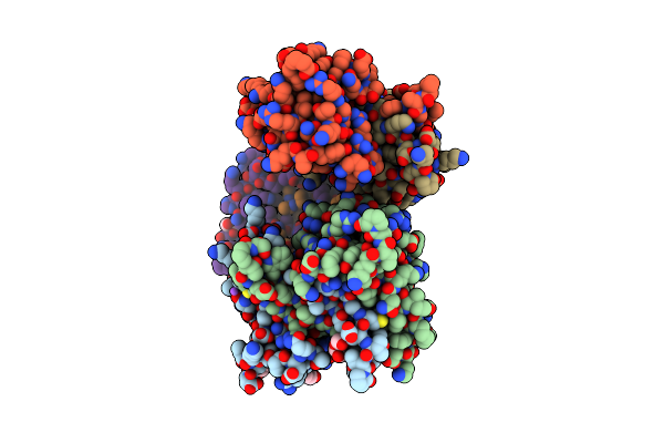
Organism: Severe acute respiratory syndrome coronavirus 2
Method: X-RAY DIFFRACTION Resolution:1.85 Å Release Date: 2022-06-22 Classification: RNA BINDING PROTEIN, Viral protein Ligands: GOL, ACT |
 |
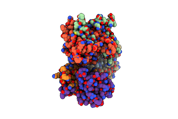
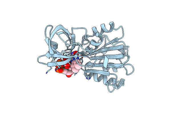
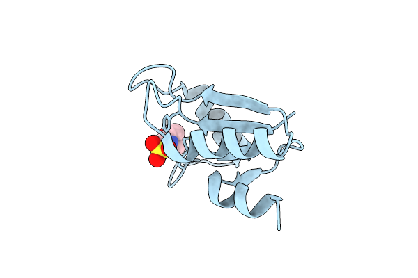
Crystal Structure Of Sars-Cov-2 Nucleocapsid Protein C-Terminal Domain Complexed With Chicoric Acid
Organism: Severe acute respiratory syndrome coronavirus 2
Method: X-RAY DIFFRACTION Resolution:1.73 Å Release Date: 2022-06-22 Classification: RNA BINDING PROTEIN/Viral Protein Ligands: GKP, PEG, GOL, NA, CL |
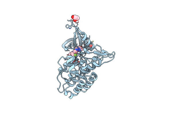 |
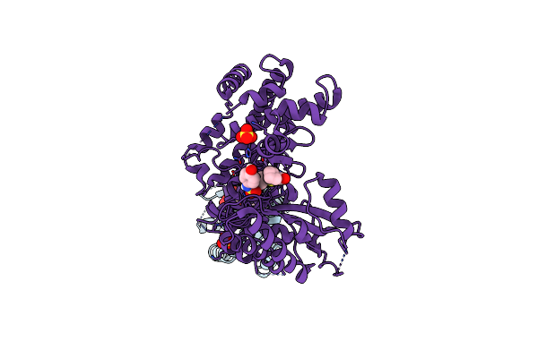
Tgf-Beta Receptor Type 1 Kinase Domain (T204D) In Complex With 6-[1-(2,2-Difluoroethyl)-4-(6-Methylpyridin-2-Yl)-1H-Imidazol-5-Yl]Imidazo[1,2-A]Pyridine
Organism: Homo sapiens
Method: X-RAY DIFFRACTION Resolution:1.83 Å Release Date: 2020-02-05 Classification: TRANSFERASE Ligands: QMY, GOL |
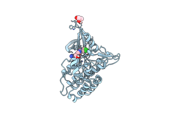 |
Tgf-Beta Receptor Type 1 Kinase Domain (T204D) In Complex With 6-[4-(3-Chloro-4-Fluorophenyl)-1-(2-Hydroxyethyl)-1H-Imidazol-5-Yl]Imidazo[1,2-B]Pyridazine-3-Carbonitrile
Organism: Homo sapiens
Method: X-RAY DIFFRACTION Resolution:1.67 Å Release Date: 2020-02-05 Classification: TRANSFERASE Ligands: QMV, GOL |
 |
Crystal Structure Of The Complex Between Ppargamma Lbd And The Ligand Am-879
Organism: Homo sapiens
Method: X-RAY DIFFRACTION Resolution:2.69 Å Release Date: 2018-02-14 Classification: NUCLEAR PROTEIN Ligands: BKY |
 |
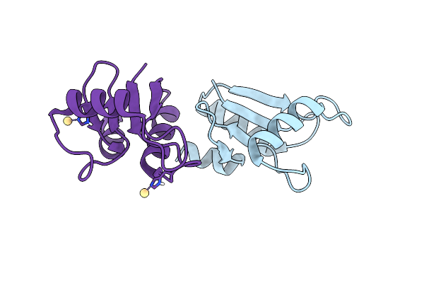
Crystal Structure Of Fat Domain Of Focal Adhesion Kinase (Fak) Bound To The Transcription Factor Mef2C
Organism: Mus musculus
Method: X-RAY DIFFRACTION Resolution:2.90 Å Release Date: 2016-07-13 Classification: Transcription, protein binding |
 |
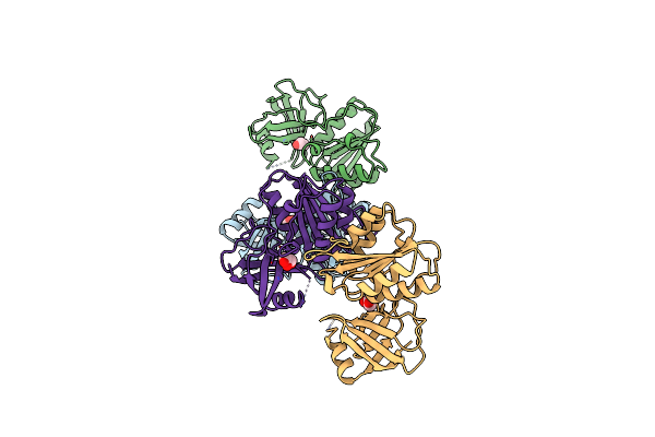
Crystal Structure Of An Engineered Construct Of Phosphatidylinositol 4 Kinase Iii Beta With A Potent And Selective Inhibitor In Complex With Gdp Loaded Rab11
Organism: Homo sapiens
Method: X-RAY DIFFRACTION Resolution:3.20 Å Release Date: 2016-02-24 Classification: Transferase/Signaling Protein Ligands: GDP, SO4, 5S8 |
 |
Organism: Escherichia coli
Method: X-RAY DIFFRACTION Resolution:2.20 Å Release Date: 1999-06-01 Classification: OXIDOREDUCTASE Ligands: GOL |
 |
Organism: Escherichia coli
Method: X-RAY DIFFRACTION Resolution:1.70 Å Release Date: 1997-09-17 Classification: FLAVOPROTEIN Ligands: FAD |
 |
Organism: Enterobacteria phage t4
Method: X-RAY DIFFRACTION Resolution:2.00 Å Release Date: 1993-10-31 Classification: ELECTRON TRANSPORT Ligands: CD |
 |
The Structure Of Oxidized Bacteriophage T4 Glutaredoxin (Thioredoxin). Refinement Of Native And Mutant Proteins
Organism: Enterobacteria phage t4
Method: X-RAY DIFFRACTION Resolution:1.45 Å Release Date: 1993-10-31 Classification: ELECTRON TRANSPORT Ligands: MES |