
Search Count: 639
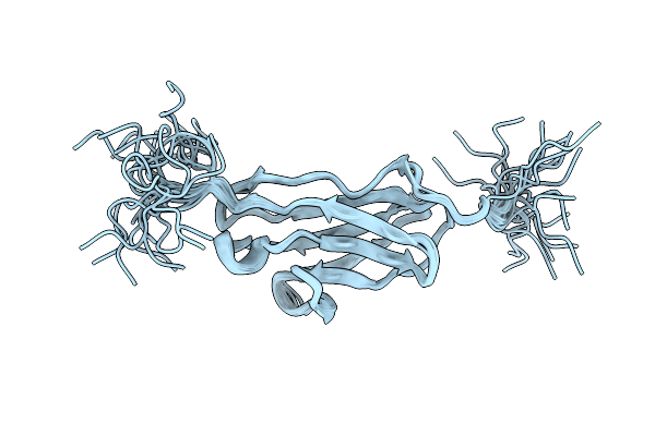 |
Solution Nmr Study Of The Titin I-Band Igi Domain I82 Reveals Conformational Dynamics
Organism: Mus musculus
Method: SOLUTION NMR Release Date: 2025-11-19 Classification: STRUCTURAL PROTEIN |
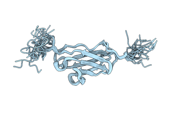 |
Solution Nmr Study Of The Titin I-Band Igi Domain I82 Reveals Conformational Dynamics
Organism: Mus musculus
Method: SOLUTION NMR Release Date: 2025-11-19 Classification: STRUCTURAL PROTEIN |
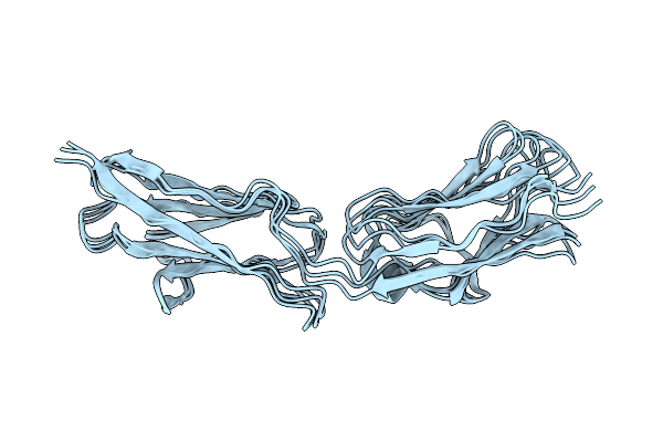 |
Organism: Mus musculus
Method: SOLUTION NMR Release Date: 2025-11-19 Classification: STRUCTURAL PROTEIN |
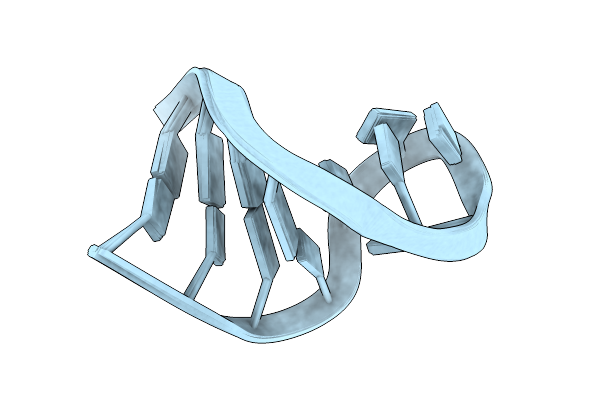 |
Nmr Structure Determination Of A Dynamic And Thermodynamically Stable Cuug Rna Tetraloop
|
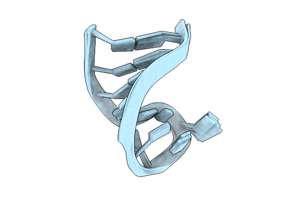 |
Nmr Structure Determination Of A Dynamic And Thermodynamically Stable Cuug Rna Tetraloop Without Restrained Loop Base Pair
|
 |
Organism: Homo sapiens
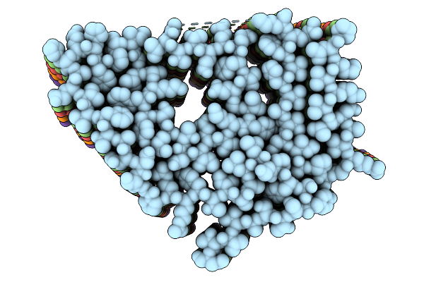
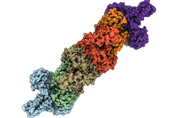
Method: ELECTRON MICROSCOPY Release Date: 2025-11-05 Classification: PROTEIN FIBRIL |
 |
Organism: Homo sapiens
Method: ELECTRON MICROSCOPY Release Date: 2025-11-05 Classification: PROTEIN FIBRIL |
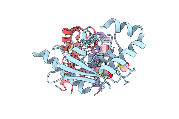 |
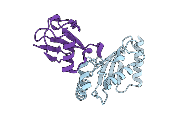
Structure Of Ubch5B In Complex With The U-Box Domain Of The E3 Ubiquitin Ligase Chip
Organism: Homo sapiens, Danio rerio
Method: X-RAY DIFFRACTION Release Date: 2025-11-05 Classification: LIGASE Ligands: BME, CL |
 |
Structure Of The Isopeptide Bond-Linked Ubch5B~Ubiquitin Conjugate Complex For An M1K/C85K Ubch5B Mutant
|
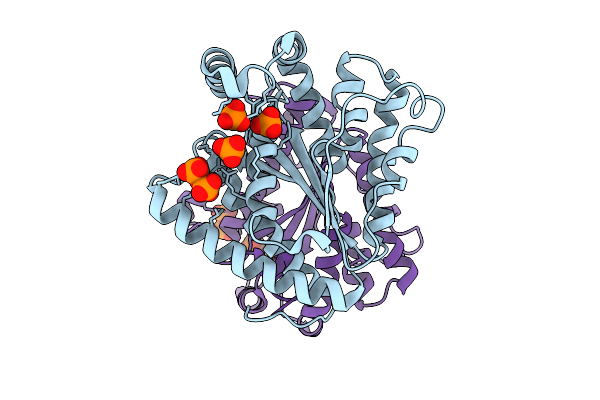 |
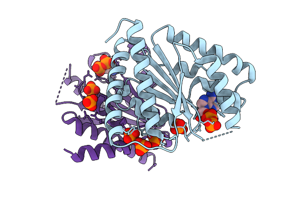
Crystal Structure Of Polyphosphate Kinase 2-Ii (Ppk2-Ii) From Bacillus Cereus Apo-Form
Organism: Bacillus cereus
Method: X-RAY DIFFRACTION Release Date: 2025-09-03 Classification: TRANSFERASE Ligands: POP, PO4 |
 |
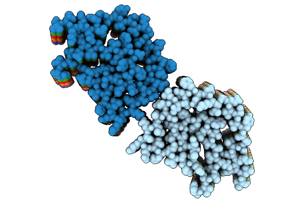
Organism: Bacillota bacterium
Method: ELECTRON MICROSCOPY Release Date: 2025-09-03 Classification: PROTEIN FIBRIL |
 |
Organism: Escherichia coli
Method: ELECTRON MICROSCOPY Release Date: 2025-08-27 Classification: RNA BINDING PROTEIN Ligands: GNP, FAD |
 |
Organism: Escherichia coli
Method: ELECTRON MICROSCOPY Release Date: 2025-08-27 Classification: RNA BINDING PROTEIN Ligands: GNP, FAD |
 |
Organism: Escherichia coli
Method: ELECTRON MICROSCOPY Release Date: 2025-08-27 Classification: RNA BINDING PROTEIN Ligands: FAD |
 |
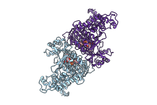
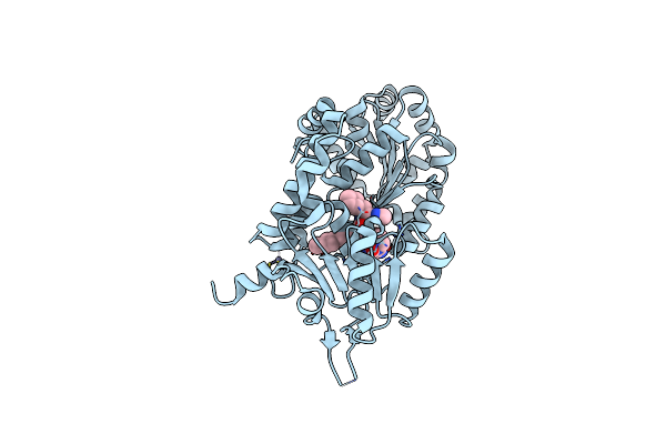
Crystal Structure Of Polyphosphate Kinase 2-Ii (Ppk2-Ii) From Lysinibacillus Fusiformis Bound To Adp (Form I)
Organism: Lysinibacillus fusiformis
Method: X-RAY DIFFRACTION Release Date: 2025-07-16 Classification: TRANSFERASE Ligands: ADP, PO4 |
 |
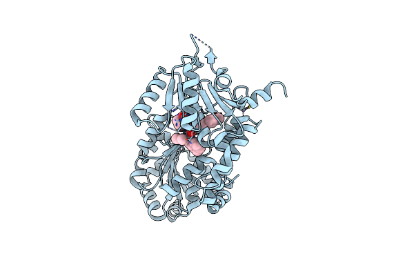
Organism: Homo sapiens
Method: X-RAY DIFFRACTION Release Date: 2025-07-09 Classification: TRANSFERASE Ligands: A1IUI |
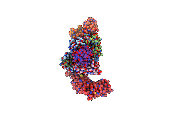 |
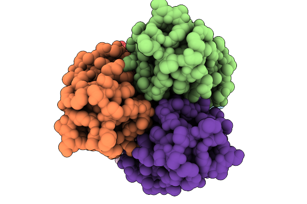
Cryoem Structure Of Mouse Garp-Ltgfbeta1 In Complex With A Fab Fragment Derived From An Activating Antibody.
Organism: Mus musculus
Method: ELECTRON MICROSCOPY Release Date: 2025-06-25 Classification: IMMUNE SYSTEM Ligands: NAG |
 |
Organism: Homo sapiens
Method: X-RAY DIFFRACTION Release Date: 2025-06-18 Classification: HYDROLASE Ligands: A1CA4, ZN |
 |
Organism: Homo sapiens
Method: X-RAY DIFFRACTION Release Date: 2025-06-18 Classification: HYDROLASE Ligands: A1CA5, ZN |
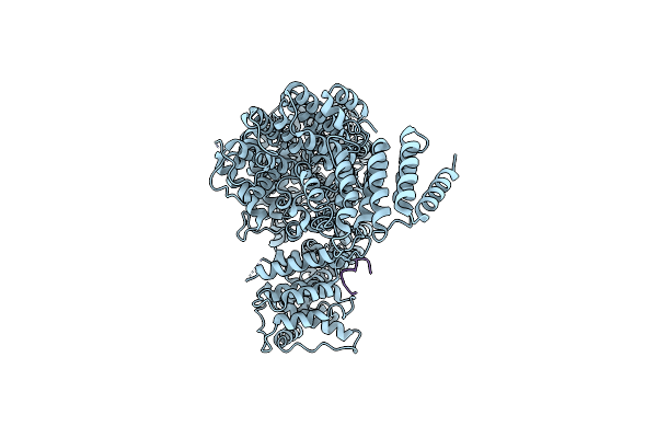 |
Cryoem Structure Of Human Mediator Subunit Med23 Complexed With Phosphorylated Elk-1 Transcription Factor
Organism: Homo sapiens
Method: ELECTRON MICROSCOPY Release Date: 2025-04-30 Classification: GENE REGULATION |