
Search Count: 24
 |
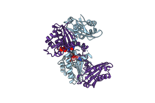
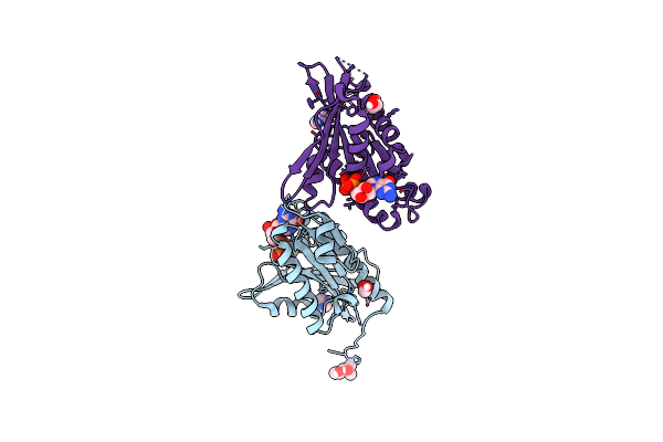
Organism: Candidatus odinarchaeota
Method: ELECTRON MICROSCOPY Release Date: 2025-07-30 Classification: CELL CYCLE Ligands: G2P |
 |
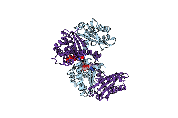
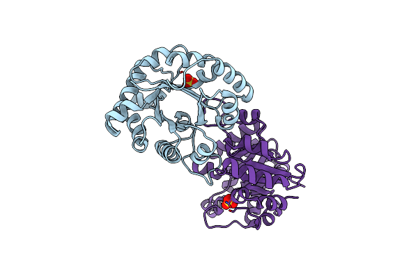
Organism: Myxococcus xanthus dk 1622
Method: X-RAY DIFFRACTION Resolution:1.89 Å Release Date: 2025-06-04 Classification: SIGNALING PROTEIN Ligands: EDO |
 |
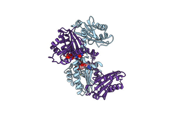
Organism: Spiroplasma melliferum kc3
Method: X-RAY DIFFRACTION Resolution:2.40 Å Release Date: 2023-09-06 Classification: CELL CYCLE Ligands: TRS, GDP |
 |
Organism: Spiroplasma melliferum kc3
Method: X-RAY DIFFRACTION Resolution:2.20 Å Release Date: 2023-08-16 Classification: CELL CYCLE Ligands: TRS, GDP, GNP |
 |
Organism: Spiroplasma melliferum kc3
Method: X-RAY DIFFRACTION Resolution:2.30 Å Release Date: 2023-08-16 Classification: CELL CYCLE Ligands: GDP, TRS |
 |
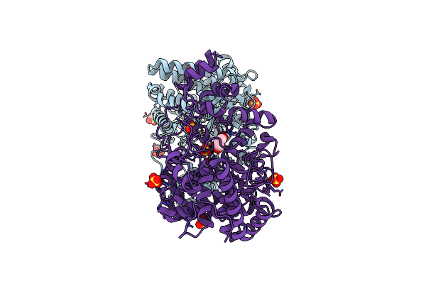
Organism: Spiroplasma citri
Method: X-RAY DIFFRACTION Resolution:2.50 Å Release Date: 2020-10-14 Classification: PROTEIN FIBRIL Ligands: ANP, PO4, MG, K |
 |
Organism: Spiroplasma citri
Method: X-RAY DIFFRACTION Resolution:2.30 Å Release Date: 2020-10-14 Classification: PROTEIN FIBRIL Ligands: ADP, MG, K |
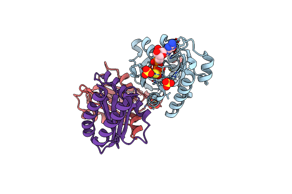 |
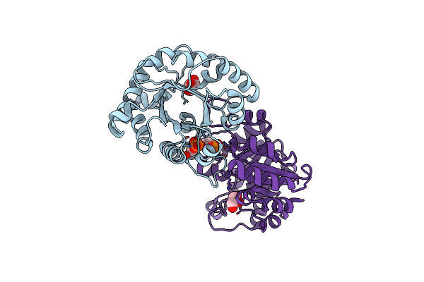
Organism: Myxococcus xanthus (strain dk 1622)
Method: X-RAY DIFFRACTION Release Date: 2019-10-16 Classification: SIGNALING PROTEIN Ligands: GSP, MG, SO4 |
 |
Structure Of Periplasmic Trehalase From Diamondback Moth Gut Bacteria Complexed With Validoxylamine
Organism: Enterobacter cloacae
Method: X-RAY DIFFRACTION Resolution:1.80 Å Release Date: 2019-01-23 Classification: HYDROLASE Ligands: SO4, VDM, GOL |
 |
Structure Of Periplasmic Trehalase From Diamondback Moth Gut Bacteria In The Apo Form
Organism: Enterobacter cloacae
Method: X-RAY DIFFRACTION Resolution:2.30 Å Release Date: 2019-01-23 Classification: HYDROLASE Ligands: SO4, GOL |
 |
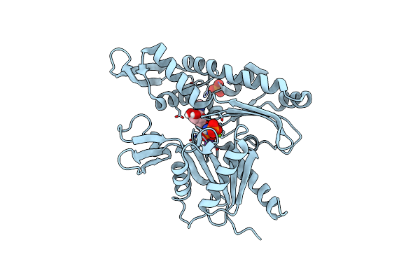
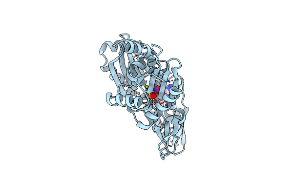
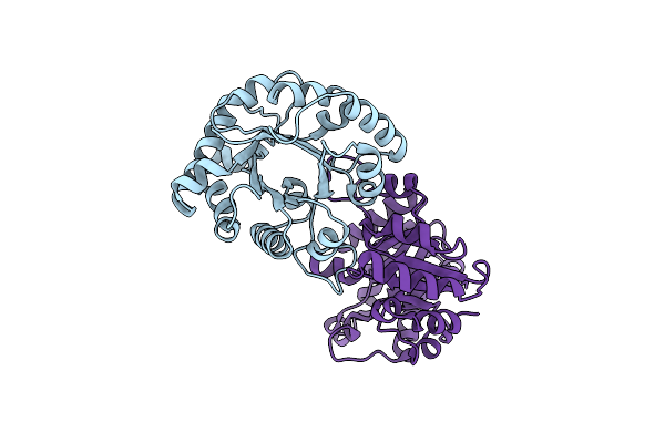
Organism: Myxococcus xanthus (strain dk 1622)
Method: X-RAY DIFFRACTION Resolution:1.35 Å Release Date: 2018-10-24 Classification: SIGNALING PROTEIN, HYDROLASE Ligands: GDP, IMD, EDO, MPD |
 |
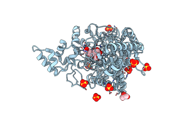
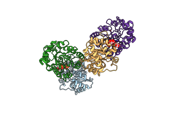
Organism: Escherichia coli
Method: ELECTRON MICROSCOPY Resolution:7.20 Å Release Date: 2012-11-21 Classification: TRANSPORT PROTEIN Ligands: ANP, MG |
 |
Organism: Escherichia coli
Method: X-RAY DIFFRACTION Resolution:2.00 Å Release Date: 2012-11-07 Classification: TRANSPORT PROTEIN Ligands: ANP, MG |
 |
Organism: Escherichia coli
Method: X-RAY DIFFRACTION Resolution:2.20 Å Release Date: 2012-11-07 Classification: TRANSPORT PROTEIN Ligands: ANP, MG |
 |
Crystal Structure Of The F96S Mutant Of Plasmodium Falciparum Triosephosphate Isomerase
Organism: Plasmodium falciparum
Method: X-RAY DIFFRACTION Resolution:1.40 Å Release Date: 2008-12-09 Classification: ISOMERASE Ligands: SO4 |
 |
Crystal Structure Of F96S Mutant Of Plasmodium Falciparum Triosephosphate Isomerase With 3- Phosphoglycerate Bound At The Dimer Interface
Organism: Plasmodium falciparum
Method: X-RAY DIFFRACTION Resolution:2.20 Å Release Date: 2008-12-09 Classification: ISOMERASE Ligands: 3PG, GOL |
 |
Crystal Structure Of The F96H Mutant Of Plasmodium Falciparum Triosephosphate Isomerase
Organism: Plasmodium falciparum
Method: X-RAY DIFFRACTION Resolution:1.70 Å Release Date: 2008-12-09 Classification: ISOMERASE |
 |
Crystal Structure Of The F96H Mutant Of Plasmodium Falciparum Triosephosphate Isomerase With 3-Phosphoglycerate Bound At The Dimer Interface
Organism: Plasmodium falciparum
Method: X-RAY DIFFRACTION Resolution:1.95 Å Release Date: 2008-12-09 Classification: ISOMERASE Ligands: 3PG |
 |
Crystal Structure Of The F96W Mutant Of Plasmodium Falciparum Triosephosphate Isomerase Complexed With 3-Phosphoglycerate
Organism: Plasmodium falciparum
Method: X-RAY DIFFRACTION Resolution:2.00 Å Release Date: 2008-12-09 Classification: ISOMERASE Ligands: 3PG |
 |
Crystal Structure Of The Plasmodium Falciparum Triosephosphate Isomerase In The Loop Closed State With 3-Phosphoglycerate Bound At The Active Site And Interface
Organism: Plasmodium falciparum
Method: X-RAY DIFFRACTION Resolution:2.25 Å Release Date: 2008-12-09 Classification: ISOMERASE Ligands: 3PG |